Difference between revisions of "Intra-Oral Radiography Interpretation - Small Animal"
Jump to navigation
Jump to search
Ggaitskell (talk | contribs) |
|||
| Line 2: | Line 2: | ||
|title = Interpretation of Intra-Oral Radiography | |title = Interpretation of Intra-Oral Radiography | ||
|categories = [[:Category:Intra-Oral Radiography|'''Intra-Oral Radiography''']] | |categories = [[:Category:Intra-Oral Radiography|'''Intra-Oral Radiography''']] | ||
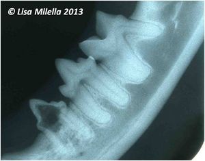
| − | |text = | + | |text = For interpretation dental radiographs should be viewed using a '''viewing box''' with minimal peripheral light and preferably using magnification. It is recommended to radiograph the '''contralateral structures for comparative purposes'''. |
|content = | |content = | ||
| Line 17: | Line 17: | ||
[[Category:Intra-Oral Radiography]] | [[Category:Intra-Oral Radiography]] | ||
| − | [[Category:To Do - | + | [[Category:To Do - Mars Check]] |
Revision as of 08:47, 2 October 2013
| ||||
| ||||

