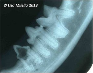
Intra-Oral Radiography Interpretation - Small Animal
| ||||
|
| ||||
| This article was written by Lisa Milella BVSc DipEVDC MRCVS. Date reviewed: 1 October 2014 |
| Endorsed by WALTHAM®, a leading authority in companion animal nutrition and wellbeing for over 50 years and the science institute for Mars Petcare. |
Error in widget FBRecommend: unable to write file /var/www/wikivet.net/extensions/Widgets/compiled_templates/wrt664539d8d02cc4_41409810 Error in widget google+: unable to write file /var/www/wikivet.net/extensions/Widgets/compiled_templates/wrt664539d8e2b764_17903457 Error in widget TwitterTweet: unable to write file /var/www/wikivet.net/extensions/Widgets/compiled_templates/wrt664539d8ef2f64_07408245
|
| WikiVet® Introduction - Help WikiVet - Report a Problem |
