Difference between revisions of "Omasum - Anatomy & Physiology"
m (Text replace - "Category:To Do - Review" to "Category:To Do - AP Review") |
|||
| Line 23: | Line 23: | ||
There are numerous small lymph nodes that are scattered in the omasal curvatures. The lymph drains to larger atrial nodes between the cardia and the omasum, then to the cistera chyli. | There are numerous small lymph nodes that are scattered in the omasal curvatures. The lymph drains to larger atrial nodes between the cardia and the omasum, then to the cistera chyli. | ||
| + | |||
| + | ==Histology== | ||
| + | |||
| + | [[Image:Omasum Histology Sheep.jpg|thumb|right|150px|Omasum Histology (Sheep) - Copyright RVC 2008]] | ||
| + | *Keratinised stratified squamous epithelium | ||
| + | |||
| + | *Lamellae thrown into leaves | ||
| + | **Different sizes | ||
| + | **Divides the lumen into narrow and uniform recesses | ||
| + | |||
| + | *Firm | ||
| + | |||
| + | *No glands | ||
| + | |||
| + | *Papillae | ||
| + | **Most small and lenticular | ||
| + | **Some large and [[Tongue - Anatomy & Physiology#Types of Papillae|conical]] | ||
| + | |||
| + | *Cicrular tunica muscularis extends into papillae of long laminae | ||
| + | |||
| + | *Lamina muscualris extends into papillae encircling tunica muscularis | ||
| + | |||
| + | *3 smooth muscle layers in papillae so they are very motile | ||
==Species Differences== | ==Species Differences== | ||
| Line 35: | Line 58: | ||
'''Test yourself with the [[The Stomachs of the Ruminant - Anatomy & Physiology - Flashcards#The Omasum|The Omasum Flashcards]]''' | '''Test yourself with the [[The Stomachs of the Ruminant - Anatomy & Physiology - Flashcards#The Omasum|The Omasum Flashcards]]''' | ||
| − | |||
| − | |||
'''Click here for information on [[Rumen - Anatomy & Physiology|rumen - Anatomy & Physiology]]''' | '''Click here for information on [[Rumen - Anatomy & Physiology|rumen - Anatomy & Physiology]]''' | ||
Revision as of 17:54, 10 December 2010
Introduction
The omasum is the third chamber in the ruminant stomach. It lies within the intrathoracic part of the abdomen so cannot be palpated manually. Instead it is examined by ausculation. The omasum has biphasic contractions which squeeze fluid out of the food before allowing the ingesta to continue into the abomasum. Absorption of volatile fatty acids continues in the omasum.
Structure
The omasum is right of the midline. The Rumen and reticulum are located to the left and the liver and body wall to the right. It is covered by lesser omentum and is bilaterally flattened. It is located at ribs 8-11. The lower pole has an extensive connection to the fundic region of the abomasum. An omasal canal is present known as the omasal groove. The floor is smooth except for low ridges and projections around the upper opening. The opening to the reticulum is at the cranial end of the omasal canal. The opening to the abomasum is at the caudal end of the omasal canal. It is a large oval opening, partly covered by overhanging abomasal folds.
Function
A function of the omasum is absorption. Food enters omasum at second biphasic reticular contraction. There are biphasic contractions. The first contraction expels fluid by squeezing the ingesta from the omasal canal between the lamellae. The second contraction expels solids by mass contraction of the omasum. Contractions are slower than the rumenoreticular contractions (see rumination). Food is squeezed between lamellae.
Vasculature
The blood supply to the omasum includes the cranial mesenteric artery, the caudal mesenteric artery and the left gastric and left gastroepiploic arteries.
Innervation
The omasum is innervated by the dorsal vagus nerve (CN X) (most important) and the ventral vagus nerve (CN X).
Lymphatics
There are numerous small lymph nodes that are scattered in the omasal curvatures. The lymph drains to larger atrial nodes between the cardia and the omasum, then to the cistera chyli.
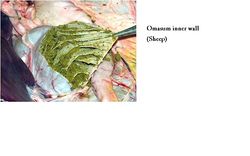
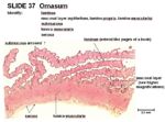
Histology
- Keratinised stratified squamous epithelium
- Lamellae thrown into leaves
- Different sizes
- Divides the lumen into narrow and uniform recesses
- Firm
- No glands
- Papillae
- Most small and lenticular
- Some large and conical
- Cicrular tunica muscularis extends into papillae of long laminae
- Lamina muscualris extends into papillae encircling tunica muscularis
- 3 smooth muscle layers in papillae so they are very motile
Species Differences
Small Ruminant
The small ruminants have a smaller omasum. The omasum is bean shaped.
Bovine
The lower pole of the omasum contacts the abdominal floor below the costal arch.
Links
Test yourself with the The Omasum Flashcards
Click here for information on rumen - Anatomy & Physiology
Click here for information on reticulum - Anatomy & Physiology
Click here for information on abomasum- Anatomy & Physiology
Video links:
Pot 52 Lateral view of the Abdomen of a young Ruminant
Pot 175 Sections of the Ruminant Stomach
Pot 47 Ovine Omasum and Abomasum
Left sided topography of the Ovine Abdomen and Thorax

