Difference between revisions of "T cells"
| Line 1: | Line 1: | ||
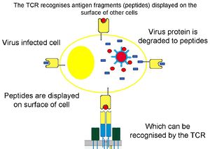
[[Image:TCR2.jpg|thumb|right|300px|T-cell receptor binds antigen fragments presented by MHC on the cell surface - B. Catchpole, RVC 2008]] | [[Image:TCR2.jpg|thumb|right|300px|T-cell receptor binds antigen fragments presented by MHC on the cell surface - B. Catchpole, RVC 2008]] | ||
| − | |||
Also known as '''T lymphocytes | Also known as '''T lymphocytes | ||
==Introduction== | ==Introduction== | ||
| − | <p>T cells are so named as they differentiate in the [[Thymus - Anatomy & Physiology|thymus]]. They are long lived and are involved in ''cell mediated immunity''. They represent 60-80% of the circulating lymphocytes and all express the markers CD2, CD3 and CD7 as well as having T cell receptors (TCR). Each T cell has 30,000 TCRs each of which is identical and recognises antigens and MHC II.</p><p>Functionally they are divided into three subsets that are distinguished by presence or absence of CD4 or CD8 markers. CD4 and CD8 cells have α/β antigen receptors while the γδ cells have the γ/δ antigens receptors.</p> | + | <p>T cells are so named as they differentiate in the [[Thymus - Anatomy & Physiology|thymus]]. They are long lived and are involved in '''cell mediated immunity'''. They represent 60-80% of the circulating lymphocytes and all express the markers CD2, CD3 and CD7 as well as having T cell receptors (TCR). Each T cell has 30,000 TCRs each of which is identical and recognises antigens and MHC II.</p><p>Functionally they are divided into three subsets that are distinguished by presence or absence of CD4 or CD8 markers. CD4 and CD8 cells have α/β antigen receptors while the γδ cells have the γ/δ antigens receptors.</p> |
* T-cell receptors are the antigen-specific receptors for T-lymphocytes | * T-cell receptors are the antigen-specific receptors for T-lymphocytes | ||
* T-cell receptors are a combination of either αβ chains or γδ chains | * T-cell receptors are a combination of either αβ chains or γδ chains | ||
| Line 12: | Line 11: | ||
* Like antibody, TCR consist of a distal domain and a membrane proximal constant domain | * Like antibody, TCR consist of a distal domain and a membrane proximal constant domain | ||
** Structurally, they look like a single arm of an antibody molecule | ** Structurally, they look like a single arm of an antibody molecule | ||
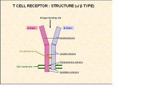
| + | [[Image:T Cell diagram 2.jpg|thumb|right|300px|T Cell - Copyright Prof Dirk Werling DrMedVet PhD MRCVS]] | ||
==Helper CD4+== | ==Helper CD4+== | ||
Revision as of 17:11, 24 September 2010
Also known as T lymphocytes
Introduction
T cells are so named as they differentiate in the thymus. They are long lived and are involved in cell mediated immunity. They represent 60-80% of the circulating lymphocytes and all express the markers CD2, CD3 and CD7 as well as having T cell receptors (TCR). Each T cell has 30,000 TCRs each of which is identical and recognises antigens and MHC II.
Functionally they are divided into three subsets that are distinguished by presence or absence of CD4 or CD8 markers. CD4 and CD8 cells have α/β antigen receptors while the γδ cells have the γ/δ antigens receptors.
- T-cell receptors are the antigen-specific receptors for T-lymphocytes
- T-cell receptors are a combination of either αβ chains or γδ chains
- One T-cell will express either αβ OR γδ TCR
- The antigen-binding site of the TCR is produced by a combination of the V domains of either 1α and 1β chain or 1γ and 1δchain
- Like antibody, the specificity of TCR is determined by the amino acid composition of the variable domains
- TCR are always linked to the cell membrane
- Like antibody, TCR consist of a distal domain and a membrane proximal constant domain
- Structurally, they look like a single arm of an antibody molecule
Helper CD4+
These T cells express the marker CD4 and are categorised into two groups, Th1 and Th2, which are distinguished by the cytokines they produce. T helper cells recognise antigens bound to MHC II complex.
- Th1 cells produce Il-2, IFN-γ and TNF-α
- Th2 cells produce Il-4, Il-5, Il-10 and Il-13
Th1 cells interact with CD8+, NK and dendritic cells and Th2 cells interact with B cells. Th1 cells are involved with the control of intracellular pathogens and Th2 cells extracellular pathogens. Il-2 produced by Th1 cells stimulates further proliferation of CD4+ cells.
Th0 populations are CD4+ cells that have yet to differentiate into Th1 or Th2 cells and they secrete Il-2, IL-4, Il-5, IFN-γ. In the presence of Il-4 they develop into Th2 cells while in the presence of Il-12 they develop into Th1 cells. In the abscence of Il-12 Th1 cells will change into Th2 cells.
Th1 cells have two populations. One that secretes IFN-γ and is short lived, and the other that doesn’t secrete IFN-γ and is long lived. The IFN-γ- cells are termed memory T cells (This relationship is not the case for Il-4+ and Il-4- Th2 cells).
For more information on T helper cells click here
Cytotoxic CD8+
These T cells express the marker CD8 and once fully mature seek and destroy target cells (infected or cancer forming cells). When the cytotoxic T cell recognises the MHC I complex on the target cell (MHC I binds to TCR) the T cell kills that cell e.g. viral peptides associate with MHC I and the CD8+ T cell recognises this and binds to the cell. Only two pathways are used by cytotoxic T cells to kill cells:
- One pathway uses the CD95 (death receptor) which triggers apoptosis in the target cell (usually other T cells)
- The other pathway uses perforins and granzymes which form pores in the target cell membrane causing cell lysis
- Perforins are structurally related to complement factor C9
- Granzymes are proteolytic enzymes that target cell nucleases and cause apoptosis
In both cases direct contact is required between the T cell and target cell, and cell killing can take several minutes.
Cytotoxic T cells secrete a pattern of cytokines similar to that of TH1 cells i.e. IFN-γ but not IL-2. IFN-γ shifts the balance of the immune response in favour of TH1 cells giving an increased level of T cell proliferation. The initiation of the immune response via cytotoxic T cells leads to the selective proliferation of cytotoxic T cells enhancing the main mechanism of killing infected cells.
γδ cells
Information on these cells is varied.
They do not express CD4 or CD8 and have γδ antigen receptors rather than α/β like other T cells. They develop in the thymus and migrate to epithelial tissues where they remain. The number present in an individual varies greatly but is generally greatest in immature ruminants and pigs.
The cells can be divided into two subsets:
- One with restricted antigen binding, that act as first line defence against invading organisms and recognises antigens bound to MHC I complex
- The other subset doesn’t require the MHC complex and this subset has a further two subsets
- One producing cytokines and chemokines (Th1 and Th2)
- The other having cytotoxic effects.

