Difference between revisions of "Tooth Development"
m (Text replace - "[[Enamel Organ#" to "[[Tooth - Anatomy & Physiology#") |
|||
| (21 intermediate revisions by 3 users not shown) | |||
| Line 1: | Line 1: | ||
| + | {{OpenPagesTop}} | ||
==Introduction== | ==Introduction== | ||
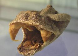
| − | [[Image:Gaboon Viper Skull.jpg|thumb|right| | + | [[Image:Gaboon Viper Skull.jpg|thumb|right|250px|Gaboon Viper - Copyright RVC]] |
| − | Teeth develop differently in different regions of the mouth in most species, a process called heterodonty. In some animals teeth develop identically in different regions of the mouth, a process called homodonty. Different species will have varying numbers of teeth and different shapes depending largely on diet. | + | Teeth develop differently in different regions of the mouth in most species, a process called '''heterodonty'''. In some animals, teeth develop identically in different regions of the mouth, a process called '''homodonty'''. Different species will have varying numbers of teeth and different shapes depending largely on their diet. Not all species possess teeth and there is huge variation in dental formulae between the species that have teeth. Teeth are mainly used for [[Mastication|mastication]] - chewing and grinding food particles, but are also used for seizing prey and tearing. The '''occlusion surface''' is where opposing teeth touch. The contact surface is where adjacent teeth touch. |
| − | |||
| − | Not all species possess teeth and there is huge variation in dental formulae between the species that have teeth. | ||
| − | |||
| − | Teeth are mainly used for [[Mastication|mastication]] - chewing and grinding food particles, but are also used for seizing prey and tearing. | ||
| − | |||
| − | The occlusion surface is where opposing teeth touch. The contact surface is where adjacent teeth touch. | ||
==Tooth Development== | ==Tooth Development== | ||
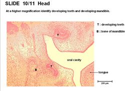
| − | [[Image:Tooth Development Histology.jpg|thumb|right| | + | [[Image:Tooth Development Histology.jpg|thumb|right|250px|Tooth Development Histology - Copyright RVC 2008]] |
| − | + | Tooth development occurs in the following stages; | |
| − | |||
| − | |||
| − | |||
| − | |||
| − | |||
| − | |||
| − | |||
| − | |||
| − | |||
| − | |||
| − | |||
| − | |||
| − | |||
| − | |||
| − | |||
| − | |||
| − | |||
| − | |||
| − | |||
| − | |||
| − | |||
| − | |||
| − | |||
| − | |||
| − | |||
| − | |||
| − | |||
| − | |||
| − | |||
| − | |||
| − | |||
| − | |||
| − | |||
| − | |||
| − | + | 1. Focal thickening of oral epithelium on the medial aspect of the [[Gingiva#Labiogingival groove|labiogingival groove]] forms the dental lamina. | |
| − | + | 2. The mesenchyme under each laminae condenses. | |
| − | + | 3. The dental lamina invaginates to form the dental bud. | |
| − | + | 4. The dental bud expands and branches to become the enamel organ. | |
| − | + | 5. The enamel organ surrounds the neural crest cell derived, dental papilla. | |
| − | + | 6. The combination of the enamel organ and dental papillae forms the deciduous tooth. | |
| − | |||
| − | + | 7. Small mass of cells bud off the dental lamina forming the primordium of the permanent tooth which continues development. | |
| − | + | 8. The inner cell layer of enamel organ (from oral epithelium) differentiates into [[Tooth - Anatomy & Physiology#Ameloblasts|ameloblasts]]. | |
| − | + | 9. Neighbouring cells in the dental papillae (from neural crest cells) differentiate into [[Tooth - Anatomy & Physiology#Odontoblasts|odontoblasts]]. | |
| − | + | 10. Dentine surrounds [[Tooth - Anatomy & Physiology#Pulp|pulp]] to produce the [[Tooth - Anatomy & Physiology#Root|root]] of the tooth. | |
| − | + | 11. Epithelial cells near the distal tooth form [[Tooth - Anatomy & Physiology#Cementoblasts|cementoblasts]], secreting [[Tooth - Anatomy & Physiology#Cementum|cementum]] around the tooth [[Tooth - Anatomy & Physiology#Root|root]]. | |
| − | + | There is a reciprocal inductive interaction between the oral epithelium and mesenchyme precursors. The mesenchyme forms the tooth, it has labile differentiative properties but stable morphogenic properties. Tooth formation starts at the [[Tooth - Anatomy & Physiology#Crown|crown]] and progresses towards the [[Tooth - Anatomy & Physiology#Root|root]]. The tooth does not acquire full length until the crown has emerged. Tooth growth is appositional. | |
| − | |||
| − | [[ | + | {{Learning |
| + | |flashcards= [[Teeth and Gingiva Flashcards]] | ||
| + | |powerpoints= [[Oral Cavity Histology resource|Histology of the oral cavity, see part 2 for developing teeth]] | ||
| + | }} | ||
| + | {{OpenPages}} | ||
[[Category:Teeth - Anatomy & Physiology]] | [[Category:Teeth - Anatomy & Physiology]] | ||
Latest revision as of 13:19, 2 November 2014
Introduction
Teeth develop differently in different regions of the mouth in most species, a process called heterodonty. In some animals, teeth develop identically in different regions of the mouth, a process called homodonty. Different species will have varying numbers of teeth and different shapes depending largely on their diet. Not all species possess teeth and there is huge variation in dental formulae between the species that have teeth. Teeth are mainly used for mastication - chewing and grinding food particles, but are also used for seizing prey and tearing. The occlusion surface is where opposing teeth touch. The contact surface is where adjacent teeth touch.
Tooth Development
Tooth development occurs in the following stages;
1. Focal thickening of oral epithelium on the medial aspect of the labiogingival groove forms the dental lamina.
2. The mesenchyme under each laminae condenses.
3. The dental lamina invaginates to form the dental bud.
4. The dental bud expands and branches to become the enamel organ.
5. The enamel organ surrounds the neural crest cell derived, dental papilla.
6. The combination of the enamel organ and dental papillae forms the deciduous tooth.
7. Small mass of cells bud off the dental lamina forming the primordium of the permanent tooth which continues development.
8. The inner cell layer of enamel organ (from oral epithelium) differentiates into ameloblasts.
9. Neighbouring cells in the dental papillae (from neural crest cells) differentiate into odontoblasts.
10. Dentine surrounds pulp to produce the root of the tooth.
11. Epithelial cells near the distal tooth form cementoblasts, secreting cementum around the tooth root.
There is a reciprocal inductive interaction between the oral epithelium and mesenchyme precursors. The mesenchyme forms the tooth, it has labile differentiative properties but stable morphogenic properties. Tooth formation starts at the crown and progresses towards the root. The tooth does not acquire full length until the crown has emerged. Tooth growth is appositional.
| Tooth Development Learning Resources | |
|---|---|
 Test your knowledge using flashcard type questions |
Teeth and Gingiva Flashcards |
 Selection of relevant PowerPoint tutorials |
Histology of the oral cavity, see part 2 for developing teeth |
Error in widget FBRecommend: unable to write file /var/www/wikivet.net/extensions/Widgets/compiled_templates/wrt662cfbd5930560_54847628 Error in widget google+: unable to write file /var/www/wikivet.net/extensions/Widgets/compiled_templates/wrt662cfbd5970d01_80053137 Error in widget TwitterTweet: unable to write file /var/www/wikivet.net/extensions/Widgets/compiled_templates/wrt662cfbd59a8307_30489115
|
| WikiVet® Introduction - Help WikiVet - Report a Problem |

