|
|
| (97 intermediate revisions by 7 users not shown) |
| Line 1: |
Line 1: |
| − | {{OpenPagesTop}} | + | {{toplink |
| − | [[Image:lambpigkidcombDM.jpg|right|thumb|250px|<small><center>A picture showing a typical lamb and pig kidney. (Courtesy of Donal McNally - University of Nottingham</center></small>]]
| + | |linkpage = Anatomy and Physiology |
| − | ==Introduction== | + | |linktext = ANATOMY & PHYSIOLOGY |
| − | The urinary system includes the [[Renal Anatomy - Anatomy & Physiology|kidneys]], the [[Ureters - Anatomy & Physiology|ureters]] which join the kidneys to the [[Urinary Bladder - Anatomy & Physiology|bladder]], the bladder itself and the [[Urethra - Anatomy & Physiology|urethras]] which permit urine collecting in the bladder to be excreted - a process termed [[Micturition - Anatomy & Physiology|micturition]]. Understanding the physiology of kidney function is key when looking at the diseases that occur in this organ, and the anatomy of all the structures within the urinary sytem is significant as a foundation to understanding the [[:Category:Urinary System - Pathology| pathology]] which affects them. The kidneys also play a vital role in the excretion of many different types of veterinary drug; [[Kidney Function and Age - Physiology|newborn and aged]] animals have altered kidney functional capacity and this is an important factor in drug excretion rates.
| + | |thispagenormal = Urinary System - Anatomy & Physiology |
| − | At a molecular level, an understanding of the principles of [[Diffusion - Physiology|diffusion]] and [[Osmosis and Filtration - Anatomy & Physiology|osmosis]] will help to understand how water and other molecules can be redistibuted between the intracellular and extracellular spaces via the [[Phospholipid Bilayer - Anatomy & Physiology|phospholipid bilayer]] that contains [[Transport Proteins - Physiology|transport proteins]] that [[Active Transport - Physiology|actively transport]] molecules such as sodium chloride.
| + | |thispagetable = Urinary System (Table) - Anatomy & Physiology |
| | + | |pagetype =Anatomy |
| | + | }} |
| | + | <br> |
| | + | |
| | + | ==Background Information== |
| | + | |
| | + | [[WikiWords#Urinary Section| Useful Definitions]] |
| | + | |
| | + | [[Transport Across Membranes - Physiology| Transport Across Membranes]] |
| | + | |
| | + | [[Introduction to Fluid Movement - Physiology |Introduction to Fluid Movement]] |
| | | | |
| | ==The Kidney== | | ==The Kidney== |
| − | The function of the kidneys is to maintain the volume and composition of plasma, regulate water, ion and pH levels, retain nutrients and excrete waste, toxins and excess electrolytes. The kidneys achieve these functions via glomerular filtration, solute reabsorption, tubular secretion, water balance and acid-base regulation.
| |
| | | | |
| − | The kidneys are paired organs which reside in the left hand side and right hand side of the dorsal abdomen respectively, and they form during [[Kidney and Urinary Tract Development - Anatomy & Physiology|development]] from the intermediate mesoderm. Their role is to [[Osmosis and Filtration - Anatomy & Physiology|filter]] the blood through the renal corpuscle; this comprises a capillary tuft known as a glomerulus which is surrounded by the Bowman's capsule within the [[:Category:Nephron|nephron]]; the movement of fluid and soluble material across these structures forms what is known as the filtrate. The filtrate is then mostly reabsorbed along the nephron until what is left comprises compounds superfluous to the requirements of the animal. Some compounds, normally fully reabsorbed, are occasionally present in the body in excess - the kidney tubules are able to respond to this excess and excrete such compounds in greater amounts. In this way the kidneys play a major role in the homeostasis of an animal. The kidneys also play a vital role in [[Fluid Movement - Physiology |total water balance]], varying their excretion of water in relation to the hydration status of the animal. Medically, the physiology of the kidneys can be manipulated using [[Diuretics Effects on Kidneys - Anatomy & Physiology|diuretic]] drugs, which inhibit the reabsorption of water from the tubules resulting in an increase in volume and therefore water loss in the urine. | + | The kidneys are paired organs which reside in the dorsal abdomen. One on the left and one on the right. Their role is to filter the blood through the glomerulus to form what is known as the filtrate. This filtrate is then on the whole reabsorbed along the nephron until what is left comprises compounds superfluous to the requirements of the organism. Some compounds, normally fully reabsorbed, are on occasion present in the body in excess. The kidney tubules are able to respond to this excess and excrete such compounds in greater amounts. This is how the kidneys play a major role in the homeostasis of the organism. The kidneys also plays a vital role in the total water balance of the organism. Varying their excretion of water in relation to the hydration status of the animal. |
| | | | |
| − | The kidneys receive 25% of cardiac output. From this they filter 20% of the plasma forming a filtrate of which all but 1% is reabsorbed. This equates to the entire circulatory volume being filtered and reabsorbed every 30 minutes. The kidneys respond dynamically to changes in blood pressure and hydration status, using several mechanisms of [[Kidney Control of Blood Pressure - Anatomy & Physiology|regulation]] including the [[Renin Angiotensin Aldosterone System| Renin-Angiotensin-Aldosterone system]] which can alter the movement of sodium chloride and water in the vascular system and extracellular spaces. | + | The kidneys receive 25% of the '''cardiac output'''. From this they filter 20% of the plasma forming a filtrate of which all but 1% is reabsorbed. This equates to all the circulatory volume being filtered and reabsorbed every 30 minutes. The functions of the kidneys are to maintain the volume and composition of plasma, regulate water, ion and pH levels, retain nutrients and excrete waste, toxins and excess electrolytes. The kidneys achieve these functions via; glomerular filtration, solute reabsorption, tubular secretion, water balance and acid-base regulation. |
| | | | |
| − | The kidneys are responsible for the production and release of two [[Kidney Endocrine Function - Anatomy & Physiology|hormones]] - Erythropoietin and Renin, which are produced in the Juxtaglomerular Cells. The kidneys also regulate the activation of vitamin D.
| + | [[Macroscopic Renal Anatomy - Anatomy & Physiology|Macroscopic Renal Anatomy]] |
| | | | |
| − | ==The Lower Urinary Tract== | + | [[The Nephron - Anatomy & Physiology|The Nephron]] |
| | + | |
| | + | [[Kidney - Blood Pressure - Physiology| The Renal Influence on Blood Pressure]] |
| | + | |
| | + | [[The Endocrine Function of the Kidney - Anatomy & Physiology|The Endocrine Function of the Kidney]] |
| | + | |
| | + | ==Lower Urinary Tract== |
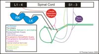
| | [[Image:sumlutshcemtri.jpg|right|thumb|200px|<small><center>A schematic summarising the structure of the lower urinary tract and it's innvervations</center></small>]] | | [[Image:sumlutshcemtri.jpg|right|thumb|200px|<small><center>A schematic summarising the structure of the lower urinary tract and it's innvervations</center></small>]] |
| − | The lower urinary tract (LUT) is the collection of organs which convey the urine formed within the kidneys to the exterior of the body. Urine is not altered in this part of the system in species other than the horse (where mucous is added) but instead the function of the LUT is to collect and store urine until enough of it is collected for release to become necessary. This gives the animal urinary continence. Three major structures make up this tract; the ureters, the bladder and the urethras which are formed from the horizontal division of the primitive hind gut in the cloacal region during [[Kidney and Urinary Tract Development - Anatomy & Physiology#Lower_Urinary_Tract|development]]. [[Urine Normal Composition|Urine]] gives valuable information to the veterinary practitioner regarding kidney function and other urinary system abnormalities such as crystals, casts and infections. | + | The lower urinary tract is the collection of organs which convey the formed urine from the kidneys to the exterior of the body. The urine is not altered in this part of the system in species other than the horse (where mucous is added) but instead its function is to collect and store the urine until enough of it is collected for release to become necessary. This gives the animal urinary continence. Three major structures make up this tract. The ureters, the bladder and the urethra. |
| | + | |
| | + | [[Ureters - Anatomy & Physiology]] |
| | + | |
| | + | [[Urinary Bladder - Anatomy & Physiology | Bladder - Anatomy & Physiology]] |
| | | | |
| − | ==Non-Mammalian Renal Systems==
| + | [[Urethra - Anatomy & Physiology | Urethra - Anatomy & Physiology]] |
| | | | |
| − | The renal anatomy and physiology of [[Exotic Urinary System - Anatomy & Physiology| fish, amphibians, birds and reptiles]] is significantly different to that of mammals.
| + | [[Process of Micturition - Anatomy & Physiology| Process of Micturition]] |
| − | Fish, for example have only a single kidney, and in physiological terms the products of excretion vary between these animal groups from that of mammals.
| |
| | | | |
| − | {{Template:Learning
| + | ==Other== |
| − | |flashcards = [[:Category:Urinary System Anatomy & Physiology Flashcards|Urinary System Flashcards]] | + | |
| − | |OVAM = [[Urinary System Vetlogic Quiz|Urinary System Quiz]]<br>[http://www.onlineveterinaryanatomy.net/content/urinary-system-and-comparative-kidneys PowerPoint presentation on the urinary system and comparative kidneys.]<br>[http://www.onlineveterinaryanatomy.net/sites/default/files/original_media/document/asset_9363_Hist.%20renal%20organs.pdf Histology of renal organs]
| + | [[Normal Composition of Urine - Anatomy & Physiology|Normal Composition of Urine]] |
| − | |Vetstream = [https://www.vetstream.com/canis/browse/Urinary Urinary disease]
| + | |
| − | }}
| + | [[Developmental Anatomy of the Kidneys and Urinary Tract - Anatomy & Physiology|Developmental Anatomy of the Kidneys and Urinary Tract]] |
| | + | |
| | + | [[Urinary Anatomy and Physiology of Exotics - Anatomy & Physiology| Exotics Anatomy & Physiology]] |
| | + | |
| | + | [[Kidney Function and Age - Physiology| Kidney Function and Age]] |
| | + | |
| | + | [[The Effects of Diuretics on the Kidneys - Anatomy & Physiology|The Effects of Diuretics on the Kidneys]] |
| | + | |
| | + | [[Urinary System - Pathology| Link to Pathology of the Urinary System]] |
| | + | |
| | + | ==Learning Resources== |
| | | | |
| − | ==References==
| + | [[Renal Flash Cards - Anatomy & Physiology| Flash Cards]] |
| | | | |
| − | Source Texts:
| + | ==Acknowledgements and Reference Material== |
| − | *{{citation|initiallast = Dyce|initialfirst = K.M|2last = Sack|2first = W.O|finallast = Wensing|finalfirst = C.J.G|year = 2002|title = Textbook of Veterinary Anatomy|ed =3rd|city = Philadelphia|pub = Saunders}}
| |
| − | *{{citation|initiallast = Sjaastad|initialfirst = O.V|2last = Hove|2first = K|finallast = Sand|finalfirst = O|year = 2004|title = Physiology of Domestic Animals|city = Oslo|pub = Scandinavian Veterinary Press}}
| |
| − | *{{citation|initiallast = Young|initialfirst = B|finallast = Heath|finalfirst = J.W|year = 2000|title = Wheater's Functional Histology: A Text and Colour Atlas|ed =4th|city = London|pub = Churchill Livingstone}}
| |
| − | *{{citation|initiallast = Hook|initialfirst = J.B|2last = Tarloff|2first = J.B|3last = Lash|3first = L.H|finallast = Goldstein|finalfirst = R.S|year = 2004|title = Toxicology of the Kidney|ed =3rd|pub = CRC Press}}
| |
| − | Websites:
| |
| − | The data found within the tables on the page entitled [[Urine Normal Composition]] was adapted from the [http://www.merckvetmanual.com/mvm/index.jsp?cfile=htm/bc/ref_00.htm Merck Veterinary Manual] online reference table entitled '''Urine Volume and Specific Gravity'''
| |
| | | | |
| − | ==Webinars==
| + | [[Acknowledgements Urinary Anatomy & Physiology|Acknowledgements]] |
| − | <rss max="10" highlight="none">https://www.thewebinarvet.com/urogential-and-reproduction/webinars/feed</rss>
| |
| | | | |
| − | [[Category:Urinary System - Anatomy & Physiology]] | + | [[Reference Material Urinary Anatomy & Physiology|Reference Material]] |
Background Information
Useful Definitions
Transport Across Membranes
Introduction to Fluid Movement
The Kidney
The kidneys are paired organs which reside in the dorsal abdomen. One on the left and one on the right. Their role is to filter the blood through the glomerulus to form what is known as the filtrate. This filtrate is then on the whole reabsorbed along the nephron until what is left comprises compounds superfluous to the requirements of the organism. Some compounds, normally fully reabsorbed, are on occasion present in the body in excess. The kidney tubules are able to respond to this excess and excrete such compounds in greater amounts. This is how the kidneys play a major role in the homeostasis of the organism. The kidneys also plays a vital role in the total water balance of the organism. Varying their excretion of water in relation to the hydration status of the animal.
The kidneys receive 25% of the cardiac output. From this they filter 20% of the plasma forming a filtrate of which all but 1% is reabsorbed. This equates to all the circulatory volume being filtered and reabsorbed every 30 minutes. The functions of the kidneys are to maintain the volume and composition of plasma, regulate water, ion and pH levels, retain nutrients and excrete waste, toxins and excess electrolytes. The kidneys achieve these functions via; glomerular filtration, solute reabsorption, tubular secretion, water balance and acid-base regulation.
Macroscopic Renal Anatomy
The Nephron
The Renal Influence on Blood Pressure
The Endocrine Function of the Kidney
Lower Urinary Tract
A schematic summarising the structure of the lower urinary tract and it's innvervations The lower urinary tract is the collection of organs which convey the formed urine from the kidneys to the exterior of the body. The urine is not altered in this part of the system in species other than the horse (where mucous is added) but instead its function is to collect and store the urine until enough of it is collected for release to become necessary. This gives the animal urinary continence. Three major structures make up this tract. The ureters, the bladder and the urethra.
Ureters - Anatomy & Physiology
Bladder - Anatomy & Physiology
Urethra - Anatomy & Physiology
Process of Micturition
Other
Normal Composition of Urine
Developmental Anatomy of the Kidneys and Urinary Tract
Exotics Anatomy & Physiology
Kidney Function and Age
The Effects of Diuretics on the Kidneys
Link to Pathology of the Urinary System
Learning Resources
Flash Cards
Acknowledgements and Reference Material
Acknowledgements
Reference Material
