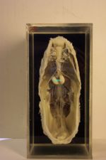
Avian Male Reproductive Tract - Anatomy & Physiology
Jump to navigation
Jump to search
Introduction
Paired reproductive tracts lie along the dorsal body wall. Each tract consists of a testis, a rudimentary epididymis and a highly convoluted deferent duct running alongside the ureter. The testes are connected to the body wall by a mesochorium. This peritoneal fold serves as an attachment for the testes and also a conduit for nerves and blood vessels.
Testes
- Bean-shaped, paired
- Lie near the cranial pole of the kidney
- Medially, they lie close to the aorta and caudal vena cava.
- Each testical suspended by a short mesochorium and surrounded medially by the abdominal air sac.
- Left tends to be larger than right in immature birds.
- Dimensions increase rapidly with sexual activity.
- In the non-breeding season, testes shrink to almost nothing and become hard to visualize.
- Dormant testes light brown/yellow in colour, turn white when sexually active.
- In some psittacine species, immature or dormant testes may appear black due to melanocytes located in the interstitium.
- Tunical Albiguinea thinner than in mammals.
- No Pampiniform plexus.
- Convoluted semniferous tubules comprised of spermatogonia (germ cells) and Sertoli cells make up the bulk of the testes. These are responsible for spermatogenesis.
- Interstitial Leydig cells produce male androgens.
- Mature spermatozoa exit into the rete testes, which connects to the cranial epididymis.
- Rete testes not present in all birds.
- Epididymis considered vestigial in birds.
- Sperm maturation occurs in the ductus deferens.
- Epididymis lies along the dorsomedial aspect of the testes.
- Spermatazoa exit the epididymis and enter the ductus deferens.
- Ductus deferens is closely associated with the Ureter in the dorsomedial midline coelom, distinguished by its zig-zag appearance.
- Under hormonal control, more convoluted in the breeding season.
- Ductus deferens enters the dorsal wall of the cloacal urodeum.
- Straightens and abruptly widens at ats junction with the cloaca.
- Structure known as the receptacle in Passerines and Budgerigars.
- Receptacle appears bean-shaped when engorged with semen.
- Birds that do not have the receptacle structure have little sperm storage capacity.
- Straightens and abruptly widens at ats junction with the cloaca.
Phallus
- Most birds lack a true phallus.
- Analogue of the mammalian penis.
- Consists of a small median tubercle flanked by a pair of large, lateral phallic bodies.
- When present, the avian phallus is soley reproductive and becomes engorged by lymph fluid instead of blood during erection.
- Owing to the lack of accessory sex glands, avian semen has low volume.
- Some lymph may contribute to the seminal fluid.
- Sperm remains viable in the female tract for much longer than in mammals.
- May survive for 5-6 days.
Absence of Phallus
- Psittacines, Passerines, Pidgeons and birds of prey all have no phallus.
- Copulate by transferring semen from the everted Cloaca directly into the female oviduct.
Non-Protrusible Phallus
- Rudimentary non-protrusible phallus is seen in male Turkeys and Chickens.
- Lies on the ventral lip of the vent.
- Consists of a small medial tubercle intimately associated on each side with lymphatic folds and vessels.
- When erect with lymph, the phallus develops a median groove.
- Median groove permits passage of ejaculate down into the everted female oviduct.
Protrusible Phallus
- Ratites and Anseriformes
- Elongated, capable of true intromission into the female cloaca.
- Distal end lies enclosed in a cavity on the floor of the cloaca and becomes engorged with lymphatic fluid.
- Anseriformes have a curved, fibrous phallus that conveys semen via a spiral groove.
Accessory Sex Organs
The male avian has no accessory sex glands that are seen in mammals. Instead, they have accessory reproductive organs:
- Paracloacal Vascular Bodies
- Dorsal Procotodeal Gland
- Lymphatic Folds
These are either in proximity to, or are an integral part of the cloaca.
Paracloacal Vascular Bodies
- Found alongside the receptacle of the vas deferens.
- Contribute to the lymphatic erection of the either cloacal or phallic tissue.
- Release a lymph-like transparent transudate when engorged.
Dorsal Procotodeal Gland
- Found on the dorsal proctodeum.
- Develop to varying degrees.
- Undergo hypertrophy in response to steroid sex hormones.
Lymphatic Folds
- Found within the wall of the procotodeum.
- Contribute to the lymphatic erection of the either cloacal or phallic tissue.
- Release a lymph-like transparent transudate when engorged.
