Cheek Teeth Disorders - Donkey
Retained deciduous teeth
Retention of the remnants of the deciduous (milk) cheek teeth or ‘caps’ can occur in donkeys between two and five years of age. The caps are normally shed at 2½, 3 and 4 years respectively in the first (06s), second (07s) and third (08s) cheek teeth, but there can be much normal variation between individual donkeys in the time that they shed their caps.
If caps are very loose or just partially retained by attachment to the gums, they may cause short-term oral pain. Affected donkeys may show headshaking, quidding, interference with the bit, and occasionally loss of appetite for a couple of days. Such signs in this age group (two- to five-year-old donkeys) warrant a careful examination of the front three cheek teeth for evidence of loose caps. If present, they may be removed using a specialised ‘cap extractor’ or a long slim-bladed instrument. Both sides of the mouth should be compared to assess if the corresponding, contra-lateral cap has already shed; if not, it can also be extracted.
The retention of caps may predispose to delayed eruption of the underlying permanent cheek tooth and the development of eruption cysts (three- or four-year-old ‘bumps’) on the mandible under the developing apices (‘root area’) of the permanent cheek teeth. The presence of very enlarged eruption cysts, especially if unilateral, should prompt a referral to a veterinary surgeon for a thorough oral and, if necessary, radiographic examination for the presence of retained deciduous cheek teeth or apical infection.
The practice of methodically removing caps at set ages in donkeys will result in the premature removal of some deciduous cheek teeth. Once the deciduous tooth is removed, the underlying fleshy dental sac covering the developing permanent cheek tooth is exposed and is then quickly destroyed while chewing food. This will lead to loss of blood supply to the infundibula (‘cups’) of the upper cheek teeth where active cement deposition is often still occurring and may possibly result in cemental hypoplasia and later dental infections. In conclusion, caps should not be removed until they become loose or the opposite one has been shed.
Rostral positioning of the upper cheek teeth
A common dental abnormality in donkeys is a rostral (forward) positioning of the upper cheek teeth arcades relative to their mandibular (lower) counterparts. In many cases this abnormality is associated with the presence of ‘parrot mouth’. Because the upper and lower cheek teeth rows are not in full contact in this condition, this leads to the development of obvious localised dental overgrowths (‘hooks’) on the rostral aspects of the first upper cheek teeth (106 and 206) that may interfere with the bit. If small, these hooks can be rasped level. If large, especially in a younger donkey, power tools may be used to reduce them. Similar overgrowths on the caudal aspect of the last (sixth) lower cheek teeth (311 and 411) are frequently undetected and can lacerate the adjacent oral mucosa during mastication. The presence of ‘parrot mouth’ and of hooks on the first upper cheek teeth should always prompt a thorough examination of the caudal lower cheek teeth.
Sedation and use of a gag is often necessary to remove the lower 11s (sixth mandibular cheek teeth) hooks and so this will need to be performed by a veterinary surgeon. There is very little room behind the lower 11s and the vertical ramus of the mandible (the upright part of the lower jaw bone) and it is easy to damage this bone when rasping vigorously.
‘Molar cutters’, cold chisels, specialised percussion guillotines which encircle this caudal hook or, most satisfactorily, rotary mechanical grinders, are used to remove these overgrowths. There is a tendency at present away from using shears, which can fracture the tooth and allow pulp exposure to occur. Such damage is most likely to occur in donkeys under ten years old, especially if they have a marked upward (dorsal) curvature of the back occlusal surface of their cheek teeth (i.e. a marked ‘Curve of Spee’). This dental fracture and pulp exposure can lead to septic infection of the mandibular and pharyngeal areas in some donkeys unless appropriate antibiotics are administered. There have been many documented cases where life-threatening pharyngeal infections have been caused by lay equine dentists attempting to remove caudal ‘hooks’, resulting in significant economic and legal loss. Grinding down such overgrowths with specialised power tools in sedated donkeys is the safest technique.
Disorders of wear
See the Dental Overgrowths section.
Smooth mouth
Smooth mouth, i.e. absence of enamel on the occlusal surface of cheek teeth, is common in old donkeys. In younger donkeys smooth mouth can result from absent or short infundibula in the upper cheek teeth, or reduced enamel infolding in the lower cheek teeth, due to increased physiological wear of the occlusal surface – that is normally around 1.5 to 2 mm per annum. At that rate of wear, most cheek teeth will be worn away in 35 to 40 years.
Enamel ridges, being much harder than the surrounding dentine or cementum, are proud on the occlusal surface. As such, feed is masticated efficiently as it is gripped and then crushed between the two opposing irregular occlusal surfaces. An irregular occlusal table also increases the occlusal surface area of each individual tooth which also increases the efficiency of each grinding stroke. Excessive rasping, especially with power tools, removes these ridges, resulting in flat, smooth tables with resultant less efficient feed utilisation. Within a few months, however, the occlusal table will again develop proud enamel ridges and return to more efficient food mastication. However, continual excessive rasping will speed up wear, and this will effectively shorten the functional lifespan of the cheek teeth. Once all enamel has been worn away in the crown (clinical and reserve), the remaining dentine and cementum are worn away at a much faster rate. Later the individual roots will become exposed and will display the characteristic hypercementosis of aged teeth.
Diastema

The occlusal surfaces of the cheek teeth are normally compressed tightly together and the six cheek teeth function as a single grinding unit. This is achieved by the action of the angled first (Triadan 06s) and last two (Triadan 10s and 11s) cheek teeth compressing together the occlusal aspect of all six cheek teeth. The cheek teeth taper slightly from crown to apex and, even with age, the progressively smaller reserve crowns usually remain tightly compressed at the occlusal surface. However, if spaces (which are termed ‘diastema’ and are often 2 to 5 mm wide) develop between the teeth, interdental food impaction will occur, especially if the diastemata are narrow on the occlusal surface and wider more apically, such as in ‘valve’ diastema.
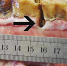
The massive and prolonged forces of mastication in the donkey will cause progressively deeper food impaction, and secondary periodontal disease will result. This disorder will be recognised by finding food fibres packed in small spaces between the cheek teeth, especially the back lower teeth, and the dental technician should refer such cases to a veterinary surgeon. Radiography, and even endoscopic examination of the mouth, may be required for diagnosis.
Disparity in the length of the cheek teeth rows
A disparity between the lengths of the upper and lower cheek teeth rows can result in overgrowths occurring unilaterally or bilaterally on the first and last cheek teeth, both upper and lower cheek teeth rows. Such overgrowths should be removed.
Displacement of the cheek teeth
Two different causes of cheek tooth displacement can occur in donkeys. In most cases, the displacements appear to be due to overcrowding of the dental rows during eruption, and this type of displacement is often bilateral. Rotation of the displaced tooth is also sometimes present. Gross dental overgrowths will develop on areas of the displaced tooth and its counterparts in the opposite row that are not in occlusal contact, and small diastema may be present between the displaced and normally positioned teeth.
Less commonly, developmentally displaced cheek teeth are found that have large diastemata between the displaced and adjacent normal cheek teeth. In such cases abnormal positioning of the developing tooth appears to be the cause of the displacement.
Acquired cheek teeth displacements, usually of the lower caudal cheek teeth, can develop in older donkeys. They are usually associated with lesser degrees of cheek teeth displacement and smaller overgrowths. These acquired displacements are believed to be caused by abnormal angulation of the occlusal surfaces.
Abnormally protruding areas of displaced cheek teeth and secondary overgrowths can lacerate the oral soft tissues, such as the cheeks and tongue, and cause bitting and quidding problems. In some cases, deep periodontal disease can occur due to deep food pocketing. This can lead to a secondary sinusitis (sometimes with food actually filling the sinus – termed an oro-maxillary fistula). Likewise maxillary or mandibular bone infections can occur.
Smaller abnormal protrusions or overgrowths can be removed with a rasp but larger areas will require a motorised tool for removal. If very extensive food pocketing is present, or if the angle of displacement is severe, the displaced tooth may have to be extracted and these extractions are readily performed per os in the sedated donkey. An excellent response is usual for these procedures.
Supernumerary cheek teeth
The presence of more than six cheek teeth in a row have been reported in donkeys. Supernumerary teeth are usually abnormally large and irregularly shaped, as if formed from two or even three immature cheek teeth.
In horses they most commonly develop at the caudal aspect of the cheek teeth rows. Because of the irregular shape of the cheek teeth, periodontal food pocketing occurs between the supernumerary cheek teeth and the sixth cheek tooth with resultant pain, and perhaps deeper infections. Additionally, if the supernumerary teeth are in just one row, and are thus unopposed, they will later form large overgrowths.
In some cases supernumerary cheek teeth should be extracted (per os if possible). In the absence of severe periodontal disease or apical infection, removal of overgrowths is all that is required.
Periapical infections
Periapical (apical) infections of cheek teeth are a significant problem in young horses, where they are inevitably accompanied by infections of the supporting bones. Their incidence in donkeys has yet to be studied, but the authors believe them to be less frequent. When diagnosed, periapical infections require veterinary treatment. Donkey owners and lay equine dentists should be aware of the signs of dental infection and thus know when veterinary attention is required. The cause of these periapical infections in many upper cheek teeth is allegedly due to food accumulation and fermentation deep in the infundibulum (enamel cups) of these teeth. This leads to destruction of the cups’ enamel walls and then to infection of the pulp cavity. The term ‘infundibular necrosis’ has also been used to describe these lesions. Localised caries, however, are very common within these infundibula and are usually harmless.
An imbalance between wear of the cheek teeth and the formation of secondary dentine (which normally prevents the pulp cavity from becoming exposed) may also occur in donkeys. When wear exceeds secondary dentine production, this leads to exposure of and, in some cases, infection of the pulp cavity.
A bony distension of the mandible, known as an ‘eruption cyst’, may often surround the apices of the second and third mandibular cheek teeth when they erupt at around three and four years of age respectively. These erupting teeth can cause considerable focal distension and thinning of the underlying mandibular bone. Due to the age at which these swellings arrive, they are also known commonly as ‘three- or four-year-old bumps’. In the lower cheek teeth, periapical abscessation most commonly involves the second and third teeth at three to five years of age. Initially this infection is confined to the periapical region. In a minority of cases it is believed that the eruption cyst may burst through the mandible, leading to exposure and infection of the underlying tooth. Impaction, overcrowding of teeth, certain mandibular conformations and deciduous teeth retention may predispose to this disorder, which is most common in ponies. In the early stages (less than three months), the infection usually remains confined to the apex, adjacent to a probable draining sinus tract, and all the pulp cavities remain vital. At this early stage medical treatment may possibly be successful.
If allowed to progress, one or more of the pulp cavities and adjacent dental tissues may become infected. At this stage, removal of the infected pulp and adjacent infected tissues is required. Endodontic (root canal) therapy or, more usually, dental extraction, will be performed by a veterinary surgeon. Endodontic therapy (by a specialist veterinary surgeon with the donkey under general anaesthetic) has been most successful with mandibular cheek teeth periapical abscessation, provided that advanced secondary caries or significant periodontal disease are not established. Currently, however, most veterinarians prefer to extract infected cheek teeth.
References
- Dacre, I., Dixon, P. and Gosden, L. (2008) Dental problems In Svendsen, E.D., Duncan, J. and Hadrill, D. (2008) The Professional Handbook of the Donkey, 4th edition, Whittet Books, Chapter 5
|
|
This section was sponsored and content provided by THE DONKEY SANCTUARY |
|---|