Colic - Donkey
Introduction
Although closely related, there are important differences in physiology and behaviour between the donkey and the horse that influence the presenting signs of certain conditions which affect both, especially abdominal pain or ‘colic’. This section discusses the possible causes, the approach to the diagnosis and the treatment of the common causes of colic in the donkey in the UK. Most consideration will be given to the treatment of the common gastrointestinal conditions causing colic. Surgical techniques are not described and standard equine surgical textbooks should be referred to if surgery is intended.
Description
Classification of colic
Many cases of colic in equines are difficult to classify as the problem often resolves and a specific cause is not identified. Severe causes of colic are frequently identified at surgery or post-mortem. White and Edwards (1999) classified colic in horses on the basis of cause in the following ways:
• diet alterations
• anatomical predisposition
• alterations in motility
• infection
• parasites
• intestinal ulceration
• pain originating from other organs/systems or ‘false colic’
The causes of colic in the donkey can be classified under the same headings, but a familiarity with the more common causes will help the practitioner to approach the diagnosis and treatment, as the predominant causes differ from those in the horse. Proudman (1992) conducted a study of first opinion colic cases and found spasmodic or unexplained conditions to be responsible for the majority (72%) of all colic episodes in the horse. Impactive colic has been found to be responsible for the majority (58%) of all colic episodes in the donkeys at The Donkey Sanctuary. When the location of the impaction was identified, 62% were found to be at the pelvic flexure (Duffield et al, 2002). Click here to see the common causes of colic at The Donkey Sanctuary.
Diagnosis
Clinical examination and the assessment of the following parameters are essentially the same as for the horse. A thorough clinical examination will also allow detection of ‘false colics’ associated with the urinary, respiratory, cardiovascular, endocrine, reproductive and musculoskeletal systems.
History
A thorough history should always be taken. Any changes in management should be noted such as change of diet, access to an excess of fermentable food or a sudden increase in exercise. Other considerations are whether the donkey is pregnant or has a history of systemic or metabolic disease.
Clinical Signs
Assessment of pain
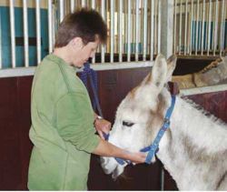
Pain is the most important criterion for assessing the presence of colic in the horse. However, donkeys show few overt signs of pain, so colic may not be identified until the donkey is in the terminal stages of the disease. A donkey in pain will often stand with its head lowered, lie down or not respond as normal. A donkey with abdominal pain is frequently described as being ‘dull’. An acute abdominal crisis may, however, present with signs similar to those seen in the horse. Abdominal ballottement and external palpation in smaller donkeys may also be useful to assess abdominal pain and distension. Dullness has been found to be the most common (90.1%) presenting sign of impactive colic followed by reduced appetite (45%) (Cox et al, 2007).
Heart rate
The normal heart rate for an adult donkey is considered to be 41 beats per minute on average. The heart rate and character of the pulse are important criteria in assessing the colic patient and also the severity of the disease. As in the horse, pain has only a relatively minor effect on the donkey’s heart rate. The rate is influenced much more by haemoconcentration and diminished venous return and by the cardiovascular response to endotoxaemia.
In the horse the elevation in pulse rate can be different, depending on the cause of the colic. Impactive colic may only cause mild elevations in pulse rate. Donkeys with impactive colic are more likely to have heart rates of 60 beats per minute or less, compared to donkeys with colic from other causes, which are more likely to have pulse rates of 60-100 beats per minute (H. Duffield's unpublished data). This may be because the impactions have little effect on circulating blood volume. It should also be considered that donkeys may cope better with dehydration and hypovolaemia due to their adaptations to arid environments.
Respiratory rate
The normal respiratory rate for the donkey is similar to that of the horse, 13-31 breaths per minute. Severe abdominal pain may increase the respiratory rate in an apparent attempt to reduce the movement of the diaphragm and chest. Pressure on the diaphragm from a grossly distended colon will also elevate the respiratory rate. An elevation in respiratory rate, accompanied by cyanosis, should be considered as evidence of very serious disease.
A donkey suffering from other respiratory diseases such as pleuropneumonia and pneumothorax may also present with signs of anorexia and depression that may be mistaken for abdominal pain.
Rectal temperature
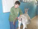
A rectal temperature should be taken routinely before the examination is performed. The normal rectal temperature of the adult donkey has been found to be slightly lower than the temperature of the adult horse with the average being 37.1°C (range 36.5-37.7°C). Elevations may occur due to physical exertion or response to infections e.g. Salmonellosis.
Mucous membrane colour and capillary refill time
The character of the mucous membranes is important for assessing the hydration of the donkey. Dehydration causes blanching of the mucous membranes and with venous congestion or endotoxin release they can become brick red and even cyanotic. The capillary refill time can be used to assess tissue perfusion and cardiovascular performance.
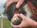
In the donkey the oral mucous membranes are normally pale pink but with less of a yellow tinge in comparison to the horse. A degree of pallor may sometimes be regarded as normal in the more elderly donkey. Gingivitis due to severe dental disease may also alter the colour of the gums. In these cases other mucous membranes may be more useful for diagnostic purposes.
Musculo-skeletal system examination
Donkeys in the UK frequently suffer from laminitis which, if severe, may present as prolonged recumbency, altered stance, depression and anorexia that may be mistaken for signs of abdominal pain. The presence of a white line abscess may also present with similar signs. The gait should be assessed and the feet examined for increased digital pulses and solar pain. (See foot diseases).
Abdominal auscultation
The character of normal borborygmi in the donkey is essentially similar to that found on examination of the horse. Due to the smaller size of the donkey, the separation of the abdomen into ‘quadrants’ may be less useful. Increased borborygmi may indicate spasmodic colic as it does in the horse, but it is more common in the donkey with abdominal pain to find a reduction or absence of borborygmi due to intestinal impactions. Reduced or absent borborygmi have been recorded in 76% of donkeys with impactions (Duffield et al, 2002).
Appetite
Appetite should be assessed in any donkey initially presenting to the practitioner as dull. This may be the only sign of abdominal pain shown. If the donkey has been anorexic for some time, secondary hyperlipaemia may have developed. A blood sample should therefore be taken for triglyceride measurement.
Rectal examination
A rectal examination should always be carried out. This is an equally important part of the clinical examination in the donkey as it is in the horse and should not be shied away from by the practitioner simply because of the size of the donkey. Plenty of lubricant, adequate restraint, careful introduction of the hand and checking the rectal sleeve for blood will help to avoid problems. Alpha-2 agonists may be used to facilitate rectal examination but are rarely necessary and may interfere with gut motility.
The faecal output and character of the faeces should be assessed. Donkeys with large intestinal impactions may have little or no faecal output. Hyperlipaemic donkeys frequently have dry, mucus covered faecal balls and the dry rectal mucosa is more prone to tearing. Coprological examinations can be carried out as a possible indicator of problems. Faecal samples may also be cultured for organisms such as Salmonella.
Displacement or distension of the intestines should be assessed. The practitioner should try to decide if the distension is due to ingesta, fluid or gas. Even if it is not possible to palpate the abdomen thoroughly, abnormal structures may be palpable at the pelvic inlet. 53% of all impactions have been found to be at the pelvic flexure (Cox et al, 2007).
Normal structures which may be palpable include the following, although this is not always the case:
• The spleen
• The left kidney
• The nephrosplenic ligament
• The caudal end of the caecum
• The pelvic flexure of the large colon and extending away from it the left ventral colon
• The small colon containing faecal balls
• A distended bladder
• The inguinal canals in the male
• The ovaries and uterus in the female
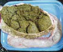
Possible abnormal findings on rectal examination are as follows:
• Distended loops of small intestine caused by obstruction or ileus of the small intestine
• A tympanitic or impacted caecum in or towards the right hand side of the pelvic inlet
• An impacted large colon either on or to one side of the pelvic floor
• Variable amounts of left colon draped over the nephrosplenic ligament if a left dorsal displacement (nephrosplenic entrapment) has occurred
• Distended large colon lying in front of the pelvic canal, caudal to the caecum if a right dorsal displacement has occurred
• Bladder stones
• Periparturient problems and other abnormalities in the uterus and ovaries
• Intra-abdominal tumours
Some large colon displacements or torsions prevent the thorough palpation of the caudal abdomen due to the small size of the donkey.
Rectal examination enables the cases that require surgical intervention to be identified. The decision to go ahead with surgical treatment in the geriatric donkey will also involve the consideration of other factors that are discussed later.
Gastric reflux
Gastric reflux should be assessed by the passing of a nasogastric tube. A small diameter tube (e.g. 9-11 mm) is recommended to avoid nasal mucosal haemorrhage. Foal tubes may be used in smaller donkeys but the practitioner should be cautious when assessing the presence of gastric reflux through a narrow diameter tube. The assessment of reflux is of diagnostic value and will also alleviate pain if the stomach is very distended. Gastric reflux greater than two litres in the horse, therefore proportionately less in the donkey, is considered significant, and usually indicates a primary disorder located in the stomach or intestine. However ileus, grass sickness and peritonitis may also prevent gastric emptying.
Abdominocentesis
Abdominocentesis and the analysis of peritoneal fluid reflects the changes that occur in tissues and organs within the abdomen. In the colic patient it can be used to determine the severity of the disease. In principle the taking of the sample is the same as it would be in the horse. However a sample may be more difficult to obtain in the donkey due to the large intra-abdominal fat pad above the linea alba. A 10 cm needle or teat cannula may be required to reach peritoneal fluid.
Examination of the oral cavity
Examination of the mouth and teeth, preferably using a Hausmann’s gag, should be carried out. In the geriatric donkey with few or no incisor teeth the use of diastema plates is preferable. The presence of dental disease is an important factor when decision-making and considering the prognosis. The significance of dental problems is explained below (see Decision-making).
Laboratory Tests
Blood tests
A normal range for biochemical and haematological parameters for the healthy donkey has been established by The Donkey Sanctuary. Complete blood count and biochemical analysis may allow the following abnormalities to be diagnosed:
- Hyperlipaemia
- Triglyceride assay may not be included in a routine screen and may need to be specifically requested
- If biochemical analysis is not available immediately then a visual assessment of the triglyceride level can be made. A blood sample should be taken in a tube without anticoagulant. When the clot retracts the serum will appear cloudy if the donkey is severely hyperlipaemic
- Hepatopathy
- Nephropathy
- Anaemia
- Immune responses to infection
- Myopathy
- Hyopalbuminaemia
Conditions affecting the liver, kidneys and pancreas may be causing the abdominal pain, i.e. ‘false colic’. If these occur concurrently with a gastrointestinal crisis then the blood results may influence the prognosis. In horses with colic, packed cell volume (PCV) and total plasma protein are used to assess hydration of the patient. The reliability of these parameters for assessing dehydration in the donkey has not yet been evaluated. It should be noted that protein may also be lost into the abdomen if peritonitis is present and the presence of hypoproteinaemia is not uncommon in the geriatric donkey. Ideally acid base balance should also be monitored in the colic patient to allow the correct choice of fluid replacement.
Decision-making
Following the physical examination, the practitioner will need to decide which course of treatment, if any, to follow. Surgery may be the only viable treatment option with certain cases of colic, whereas in other cases, medical treatment may be the initial choice, e.g. with large intestinal impactive conditions. If the exact diagnosis is not obtained, which is often the case, then the following points are taken into consideration when deciding if surgical intervention is required.
• Palpation of distended intestines
• Presence of abnormal peritoneal fluid
• The reduction or absence of intestinal sounds
• Significant amounts of gastric reflux if there is a proximal gastrointestinal obstruction
• Severe abdominal pain
The duration of the colic episode is also important. As mentioned earlier, the donkey with colic is often not identified until it is in the terminal stages of disease. The patient may therefore present with a progressively increasing heart rate, congestion of the mucous membranes and possibly an increased triglyceride level due to a period of anorexia or inappetance. These factors all make the donkey a less favourable surgical candidate.
When considering surgery in the geriatric donkey in the early stages of an abdominal crisis, additional factors have to be considered before severe circulatory compromise has occurred. If there is severe dental disease present this may have been a contributing factor to the development of the impaction and may impair recovery post surgery, due to the decreased ability to masticate long fibres. Dental disease may also increase the likelihood of the recurrence of any impaction. Concurrent conditions, such as liver disease, renal disease, pancreatitis, anaemia, immune responses to infection, or hypoalbuminaemia, will all heavily influence the decision to go to surgery. The presence of chronic musculo-skeletal conditions such as laminitis and arthritis should also be considered, as medical or surgical treatment of the colic may exacerbate such conditions.
Treatment
The primary aims of medical treatment of colic are to relieve pain, correct dehydration and restore the normal passage of ingesta. Some further discussion is given to the treatment of the impactions, but treatment of other types of colic should follow the same protocols as in the horse.
Pain relief
Relief of gastric distension using nasogastric intubation will help to relieve pain. NSAIDs (non-steroidal anti-inflammatory drugs), especially flunixin meglumine, are commonly used to control the pain associated with colic. The potential for gastric mucosal problems and renal toxicity should also be considered when administering NSAIDs. Spasmolytics may also relieve the signs of colic.
Correcting dehydration
Rapid infusion of a balanced electrolyte solution via an intravenous catheter will restore circulating blood volume and improve tissue perfusion. It will also help to correct shock caused by the absorption of endotoxins through a compromised intestinal wall. Intravenous infusion of fluids is useful when rehydration via the oral route is inappropriate, i.e. when there is gastric reflux, ileus or an intestinal obstruction. If perfusion is very poor, hypertonic saline may help in the short term but must be followed up quickly by large volumes of isotonic fluids.
Rehydration may also be achieved by the administration of oral fluids via a nasogastric tube. This method is only appropriate when no gastric reflux/intestinal obstruction is present. The administration of oral fluids via a nasogastric tube should be stopped if pain is caused, although this may be difficult to detect in the stoical donkey.
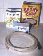
Feeding
When a lesion requiring surgical corrrection has been diagnosed, feeding may be contra-indicated until post surgery. However, a source of nutrition is needed as soon as possible in order to maintain a positive energy balance and prevent the development of hyperlipaemia. Rectal findings should be monitored, and in horses with caecal or colon impactions it is believed that feed should only be given once the impaction is no longer palpable. However, in H. Duffield's experience, if the impacted donkey wants to eat and there are normal borborygmi present and a good faecal output, then small amounts of high calorie feed with a high water content may be useful to prevent hyperlipaemia. Mineral oil or glucose powder may also be added to the feeds to avoid using a nasogastric tube.
Blood parameters need to be monitored closely, especially glucose levels which need checking every four hours.
Turning an impacted donkey out into the field to eat fresh grass and move around may also encourage the passage of ingesta. Long fibres such as hay and straw should be restricted until the normal transit of ingesta has been completely re-established and should be reintroduced gradually.
If oral nutrition is not possible due to ileus, then partial or total parenteral nutrition can be used to supply energy. The Donkey Sanctuary has little experience with this kind of therapy. Hospitalisation is required for this treatment and in certain cases the movement of an anorexic, geriatric donkey to a hospital may not be indicated.
Nursing Care
Provide the donkey with a deep bed, as it may go down and start rolling if its condition deteriorates. Companions should be separated, but kept in sight. The patient can then be left on intravenous fluids, with faecal output and food intake monitored. It may be necessary to withhold food.
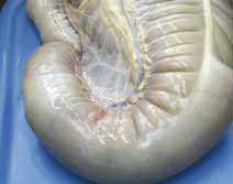
Colonic impaction is a common cause of colic in donkeys associated with dental problems, diet changes and hyperlipaemia.
Impactive conditions
Gastric ulceration
Pancreatitis
Literature Search
Use these links to find recent scientific publications via CAB Abstracts (log in required unless accessing from a subscribing organisation).
Colic in donkeys publications
References
- Duffield, H. (2008) Colic In Svendsen, E.D., Duncan, J. and Hadrill, D. (2008) The Professional Handbook of the Donkey, 4th edition, Whittet Books, Chapter 3
- Dabinett, S. (2008) Nursing care In Svendsen, E.D., Duncan, J. and Hadrill, D. (2008) The Professional Handbook of the Donkey, 4th edition, Whittet Books, Chapter 18
- Cox, R., Proudman, C., Burden, F., Trawford, A., Gosden, L., and Pinchbeck, G. (2007). ‘A case control study to investigate risk factors for impaction colic in UK donkeys’. Proceedings of British Equine Veterinary Association Congress 2007.
- Duffield, H.F., Bell, N., and Henson, F.M.D. (2002). The Seventh International Equine Colic Research Symposium Handbook. p. 122, Manchester 14-16 July 2002.
- Edwards, G.B., White, N.A. (1999). The Handbook of Equine Colic. Butterworth-Heinemann. pp 3-4.
- Proudman, C.J. (1992). ‘A two year prospective study of equine colic in general practice’. Equine Veterinary Journal. 24(2). pp 90-93.
|
|
This section was sponsored and content provided by THE DONKEY SANCTUARY |
|---|
