Endodontic Treatment
Introduction
Endodontics refers to the treatment of the pulp of the tooth (Endo: inside; -dontic: tooth). There are three pulpal treatments, each of which has specific indications. These are:
- Pulp capping
- Partial pulpectomy with direct pulp capping
- Root canal therapy
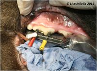
Conventional root canal therapy is the most commonly indicated type of endodontic treatment. It involves total removal of pulp tissue, i.e. total pulpectomy, cleaning and filling of the root canal, followed by tooth restoration.
Indications
Endodontics is indicated when there is, or may be, irreversible pulp pathology (e.g. generalized pulpitis or pulp necrosis, often in combination with periapical involvement). Such cases include fractures of the crown when the pulp is exposed, non-vital teeth, dental caries and iatrogenic pulp exposure in cases of crown shortening for malocclusion problems. In all of the above cases, extraction may be an option. Although endodontic treatment is often less traumatic and conserves strategically important teeth for both cosmetic and functional purposes it does require referral to a veterinary dental specialist for treatment.
Contraindications
Endodontic treatment requires long term radiographic monitoring, therefore a patient who is a high anaesthetic risk may not be a suitable candidate. In some cases, root canal treatment may also involve a longer surgery time, in which case, extraction is the preferable treatment for these patients.
Objectives
The objectives of conventional root canal therapy are:
- To clean and disinfect the pulp chamber and root canal(s).
- To fill the root canal(s) with a non-irritant, antibacterial material, thus sealing the apex.
- To close the access and exposure sites with a suitable restorative material.
- Root Canal Therapy
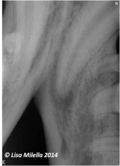
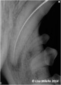
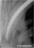
The whole procedure is performed under general anesthesia and under strict radiographic control. It is time-consuming, as each step needs to be performed with meticulous detail to ensure a successful outcome. The outcome of conventional root canal therapy should be monitored radiographically for 6–12 months postoperatively, then ideally every year thereafter.
A partial pulpectomy and direct pulp capping procedure are indicated for recent tooth crown fractures with pulp exposure in an immature tooth. An immature tooth has a thin dentine wall and an open apex, allowing a good blood supply to the pulp. Treatment is aimed at maintaining a viable pulp, as this is needed for continued root development.
- Partial Pulpectomy and Direct Pulp Capping
Complications
Failure of the endodontic treatment can occur. This is often detected radiographically. Reported success rates in dogs are 86% plus. In cases of endodontic failure, a surgical endodontic procedure can be carried out or the tooth could be extracted.
| This article was written by Lisa Milella BVSc DipEVDC MRCVS. Date reviewed: 1 October 2014 |
| Endorsed by WALTHAM®, a leading authority in companion animal nutrition and wellbeing for over 50 years and the science institute for Mars Petcare. |
Error in widget FBRecommend: unable to write file /var/www/wikivet.net/extensions/Widgets/compiled_templates/wrt699fc526ae94d5_38621624 Error in widget google+: unable to write file /var/www/wikivet.net/extensions/Widgets/compiled_templates/wrt699fc526be7833_23881326 Error in widget TwitterTweet: unable to write file /var/www/wikivet.net/extensions/Widgets/compiled_templates/wrt699fc526cd6aa0_47941677
|
| WikiVet® Introduction - Help WikiVet - Report a Problem |
