Dental Malocclusion
Introduction
By definition, malocclusion is an abnormality in the position of the teeth (normal occlusion). The clinical significance of malocclusion is that it may cause discomfort and sometimes pain to the affected animal. In some cases, it may be the direct cause of severe oral pathology.
Signalment
With the mesocephalic skull shape, the mandible is shorter and narrower than the upper jaw. In dolicocephalic breeds the upper jaw is longer than normal. The increased jaw length results in interdental spaces that are wider than normal. Brachycephalic animals have an upper jaw which is shorter than normal. A short jaw results in reduced interdental spaces with rotation and/or overlap of teeth.
Malocclusion is common in the dog, but also occurs in cats.
Malocclusion Types
Malocclusion can result from jaw length and/or width discrepancy (skeletal malocclusion), from tooth malpositioning (dental malocclusion), or a combination of both. The development of the occlusion is determined by both genetic and environmental factors. Specific genetic mechanisms regulating malocclusion are unknown. However, a polygenic mechanism is likely and explains why not all siblings in successive generations are affected by malocclusion to the same degree, if at all. With a polygenic mechanism, the severity of clinical signs is linked to the number of defective genes. The most reasonable approach to evaluate whether malocclusion is hereditary or acquired is as follows:
- Skeletal malocclusion is considered inherited unless a developmental cause can be reliably identified.
- Pure dental malocclusion, unless known to have breed or family predisposition, should be given the benefit of the doubt and is not necessarily considered to be inherited.
An outline of the more common types of malocclusion is given below.
Skeletal Malocclusion
Mandibular Prognathic Bite: In the mandibular prognathic bite, often called ‘undershot’, the mandible is longer than the maxilla and some or all of the mandibular teeth are rostral to their normal position. If the dental interlock prevents the mandible from growing rostrally to its full genetic potential, lateral or ventral bowing of the mandible may occur to accommodate the shortening in length. This results in an open bite and is characterized by increased space between the premolar cusp tips. In addition, the caudal angle of the mandible is caudal to the temporomandibular joint to accommodate the extra length of the mandible.
Mandibular prognathic (undershot) bite

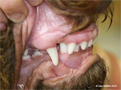
Mandibular Brachygnathic Bite: A mandibular brachygnathic bite, often called ‘overshot’, and occurs when the mandible is shorter than normal. This often results in the mandibular canine teeth contacting the palate, causing trauma.
Mandibular brachygnathic (overshot) bite

Wry Bite: A wry bite occurs if one side of the head grows more than the other side. In its mildest form a one-sided prognathic or brachygnathic bite develops. In more severe cases, a crooked head and bite develop with a deviated midline. An open bite may also develop in the incisor region so that the affected teeth are displaced vertically and do not occlude.
Wry bite
Wry bite


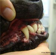
Narrow Mandible: In some animals, the mandible is too narrow with respect to the upper jaw. The result is that the lower canines impinge on the maxillary gingivae or the hard palate instead of fitting into the diastema between the upper third incisor and upper canine on either side. The animal may not be able to close its mouth and injury to the gingivae or palatal mucosa commonly occurs. In severe, untreated cases an oronasal communication may develop over time. This condition is seen in both the primary (deciduous) and permanent dentition. Persistent mandibular primary canines will further exacerbate the condition, as the permanent mandibular canines erupt medially to their primary counterparts. The incorrect dental interlock will interfere with the normal growth in width and length of the developing mandible. The condition can also be caused by persistent primary mandibular canines in a mandible of normal width.
Dental Malocclusion
Dental malocclusion is malpositioning of teeth where there is no obvious skeletal abnormality, i.e. there is no jaw length or width discrepancy.
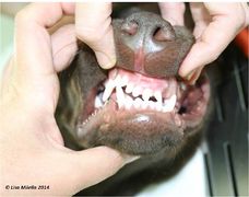
Anterior Crossbite: This is a clinical term used to describe a reverse scissor occlusion of one, several or all of the incisors. The condition can be secondary to persistent primary incisors. However, there is probably a skeletal origin as well, since affected animals often develop a mandibular prognathic bite. In other words, an anterior crossbite in an immature animal may be the first sign of a developing mandibular prognathism. The cause can either be a dental malocclusion (i.e. linguoversion of the upper incisors) or a skeletal malocclusion (i.e. mandibular prognathism or maxillary brachygnathism).
Anterior crossbite

Malocclusion of the Canine Teeth: The two most common abnormalities in canine tooth position are:
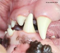
- Rostral displacement (mesioversion) of the maxillary canines (also known as lance canines). Persistent primary canines may be responsible for this condition. A breed predisposition has been reported in the Shetland sheepdog.
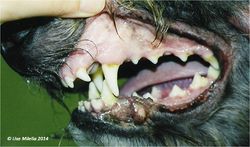
- Medial displacement of the lower canines.
Medial displacement of lower canines

Malocclusion of the Premolars and Molars: Posterior crossbite is used to describe an abnormal relationship of the carnassial teeth, seen commonly in the dolicocephalic breeds, where the normal buccolingual relationship is reversed.
Classification of Malocclusion
Class 1 Malocclusion
Also called neutroclusion, this describes a normal rostral-caudal relationship of the maxillary and mandibular dental arches but there is malposition of one or more individual teeth.
Class 2 Malocclusion
Also called mandibular distoclusion, mandibular brachygnathism or mandibular retrognathism.
In layman's terms this is referred to as an overshot jaw or a parrot mouth.
This describes an abnormal rostral-caudal relationship between the dental arches in which the mandibular arch occludes caudal to its normal position relative to the maxillary arch.
Breeds commonly affected include the Rough Collie and the Borzoi.
Class 3 Malocclusion
Also called mandibular mesioclusion, mandibular prognathism.
In layman's terms this is referred to as an undershot jaw.
This describes an abnormal rostral-caudal relationship between the dental arches in which the mandibular arch occludes rostral to its position relative to the maxillary arch.
One of the breeds most commonly affected is the Boxer.
Clinical Importance of Malocclusion
Even an otherwise sound, well cared-for dentition may show asymmetric abrasion or attrition.
Problems may occur with mastication and temporomandibular joint function.
Soft tissue trauma is a common sequel of malpositioned teeth.
Premature loss of teeth may be caused by an increased liability to periodontitis.
Owners should also be counselled with regard to the possible inheritance of the condition. Dogs with severe dental malocclusion should not be bred from.
Prevention and Treatment of Malocclusion
Prevention is always better than treatment. Early recognition of a problem is essential to avoid discomfort and pain to the animal and prevent the development of severe pathology. Malocclusion affecting the primary dentition may require interceptive orthodontics (usually extraction of the deciduous teeth). Malocclusion affecting the permanent dentition may need no treatment at all, if it is not causing the animal discomfort or any oral pathology. Malocclusion causing discomfort and pathology always needs treating.
Primary teeth involved in malocclusion should be extracted as early as possible, i.e. at 6–8 weeks of age. This will allow the maxilla and mandible to develop to their full genetic potential independently before the permanent dental interlock forms. The roots of primary teeth are longer and narrower than the roots of the permanent teeth. Extraction requires care and patience to avoid tooth fracture. It is essential not to fracture the root, as a remnant may continue to deviate the eruption pathway of the permanent tooth. Every attempt should be made to minimize the risk of iatrogenic damage to the developing permanent teeth.
The treatment options available for permanent teeth causing a traumatic malocclusion are orthodontics, tooth shortening or extraction. In many instances, tooth shortening or extraction are preferable to orthodontics on ethical grounds. Tooth shortening often requires pulpal exposure. In this situation, endodontic therapy of the shortened tooth is mandatory. Lingually displaced mandibular canines in young dogs can often be corrected by stimulating the dogs to play, as often as possible, with specific rubber toys of an appropriate size and shape.
| Dental Malocclusion Learning Resources | |
|---|---|
To reach the Vetstream content, please select |
Canis, Felis, Lapis or Equis |
Test your knowledge using flashcard type questions |
Veterinary Dentistry Q&A 09 |
References
Verstraete, F. (1999) Self-assessment colour review of veterinary dentistry Manson Publishing
| This article was expert reviewed by Lisa Milella BVSc DipEVDC MRCVS. Date reviewed: 21 August 2014 |
| Endorsed by WALTHAM®, a leading authority in companion animal nutrition and wellbeing for over 50 years and the science institute for Mars Petcare. |
Error in widget FBRecommend: unable to write file /var/www/wikivet.net/extensions/Widgets/compiled_templates/wrt69f52622ec5790_05050362 Error in widget google+: unable to write file /var/www/wikivet.net/extensions/Widgets/compiled_templates/wrt69f526230bc138_29168279 Error in widget TwitterTweet: unable to write file /var/www/wikivet.net/extensions/Widgets/compiled_templates/wrt69f526231e7af4_88012774
|
| WikiVet® Introduction - Help WikiVet - Report a Problem |