Listeria species - Overview
| Listeria | |
|---|---|
| Phylum | Firmicutes |
| Class | Bacilli |
| Order | Bacillales |
| Family | Listeriaceae |
| Genus | Listeria |
Overview
There are 6 species of Listeria bacteria. They are known as saprophytes in soil. L.monocytogenes and L.ivanovii are pathogens carried by sheep and goats and shed in faeces and milk especially during stress. They can cause septicaemia, encephalitis, abortion and endophthalmitis in ruminants. Outbreaks of Listeriosis often linked to silage feeding.
Characteristics
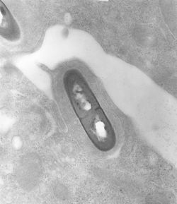
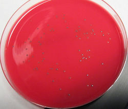
Listeria are intracellular pathogens. They are Gram positive rods with catalase positive and oxidase negative activity. They are facultative anaerobes and have tumbling motility. L.monocytogenes is haemolytic on blood agar due to a cytolytic protein, listeriolysin, and grows at range of pH values and temperatures. L.ivanovii produces a strong haemolytic zone. Listeria produces small, smooth, transparent colonies after 24 hours incubation. They are able to grow on non-enriched media.
Pathogenesis and pathogenicity
Listeria cause infection by ingestion of contaminated feed. The bacteria penetrate M cells in intestinal Peyer's patches and spread to tissues via blood and lymph. Transplacental transmission can also occur in pregnant animals. The bacteria may gain entry via breaks in oral or nasal mucosa and migrate in cranial nerves to cause neural signs. This can cause the formation of microabscesses and perivascular lymphocytic cuffs in the brainstem. L.monocytogenes can replicate within phagocytic and non-phagocytic cells, and pass between cells without being exposed to the immune system. Surface proteins known as internalins allow adherence and uptake of the bacteria into cells. Listeriolysin produced by virulent strains destroys membranes of phagocytic vacuoles, releasing the bacteria into the cytoplasm. In the cytoplasm, Listeria are motile. The bacteria can then induce formation of pseudopod projections in the cytoplasmic membrane, which are taken up with the bacteria into adjacent cells. A cell-mediated immune response is required for protection.
Diagnosis
Specimens for diagnosis should include; CSF in neural cases, cotyledons in abortion and liver, spleen and blood in septicaemia. Immunofluorescence using monoclonal antibodies can be used. Histology of brain demonstrates microabscesses and lymphocytic cuffing in the brainstem. Smears of cotyledons can be diagnostic in abortion. High protein and cell counts in CSF can be taken in neural cases and isolation on blood and MacConkey agar in septicaemia.
See here for a list of Listeria species
| This article has been peer reviewed but is awaiting expert review. If you would like to help with this, please see more information about expert reviewing. |
Error in widget FBRecommend: unable to write file /var/www/wikivet.net/extensions/Widgets/compiled_templates/wrt69a41927864544_46493374 Error in widget google+: unable to write file /var/www/wikivet.net/extensions/Widgets/compiled_templates/wrt69a419278bd9e3_04524770 Error in widget TwitterTweet: unable to write file /var/www/wikivet.net/extensions/Widgets/compiled_templates/wrt69a419279103a2_49296609
|
| WikiVet® Introduction - Help WikiVet - Report a Problem |

