Post-Mortem Change - Pathology
Jump to navigation
Jump to search
Algor Mortis
- Cooling of body after death.
- Can aid in estimating time of death.
Rigor Mortis
- Rigor mortis is a slow stiffening of the muscles caused by enzymatic activity after death.
- Affect striated muscle.
- Skeletal muscle.
- Cardiac muscle.
Process of Rigor Mortis
- There is a burst of metabolic activity as substrates are depleted on the cessation of circulation.
- Heat is liberated, giving a transient increase in temperature.
- Glycogen is broken down to lactic acid.
- The lactic acid produced is degradged no further, unlike in the living animal.
- There is a progressive decrease in muscle pH.
- Muscle oxygen and ATP is also depleted.
- There is a progressive decrease in muscle pH.
- There are two possible methods by which lowered muscle pH produces contraction:
- The acidic condtion causes Ca++ efflux from the sarcoplasmic reticulum, leading to contraction of muscle fibres.
- The acid coagulates the protein in the muscle cells, producing a kind of muscular contraction.
Onset of Rigor Mortis
Factors
- The time to onset and the degree of rigor mortis expressed by the carcass varies with a number of factors.
- If the animal has been ill and weak for some time there may be little glycogen stored in the muscles.
- Since rigor development depends on glycogen, it may a slight, transient phenomenon compared with the normal process.
- The animal may have been in a state of great muscular activity at the time of death.
- E.g.
- Being chased.
- Suffering muscle spasm, as in tetanus or strychnine poisoning.
- In this case, ante-mortem spasm may lead straight on to post-mortem rigor.
- E.g.
- Since it is dependent on enzyme activity, the onset of rigor mortis is influenced by the ambient temperature.
- If the animal has been ill and weak for some time there may be little glycogen stored in the muscles.
Time Course
- Under normal conditions, rigor develops about 2-8 hours after death, and lasts for a further 36 to 72 hours.
- The animal dying today would go into full rigor today, and remain in rigor tomorrow.
- Rigor starts to wear off on the third day.
- Due to further autolysis breaking down the coagulated protein in the muscle fibres.
- If rigor is broken forcibly, e.g. by flexing a limb when it is in rigor, it does not recur.
- This is because no further coagulation contraction is possible.
Pattern in Body
- Rigor mortis tends to occur first in the muscles that are normally most active.
- The heart is first to be affected.
- Causes strong contraction of the left ventricle, which is sufficient to expel the blood it contained.
- The diaphragm contracts next.
- The head and neck musculature then follows.
- The jaw muscles are rich in glycogen, so it may be very difficult to prise open the jaw.
- Rigor of the muscles of the eye mean the eye is retracted into the socket and appears sunken.
- It may also appear cloudy since the cornea dries out and appears opaque.
- The changes then spread to the extremities; first the forelimbs and then the hindlimbs.
- Affects both the extensor and flexor muscles of the limbs.
- Due to the relative strengths of these muscles, the net result is towards extension of the stiffening limbs.
- In an unconfined space, the animal is seen lying on its side with its legs stuck out like posts.
- Due to the relative strengths of these muscles, the net result is towards extension of the stiffening limbs.
- Affects both the extensor and flexor muscles of the limbs.
- The heart is first to be affected.
- There appears to be a wave of rigor passing down the body beginning at the jaw and ending with the hindlimbs.
- Rigor tends to wear off in the same way as it begins.
- Those muscles showing rigor first tending to lose it first.
Interpretation of Rigor Mortis
- If rigor is well developed immediately after death, this may indicate :
- An increased activity just before death.
- Some poisoning or disease.
- If rigor comes on imperfectly, this may indicate that the animal was weak or ill before it died.
- Failure of the left ventricle to expel its blood before indicates that the heart did not undergo proper rigor.
- Seen quite often in toxaemia.
Post-Mortem Clotting of Blood
- Blood coagulates in the vessels - seen in:
- Large arteries
- The right ventricle, since it has less contractile power in rigor mortis than the left ventricle.
- Quite distinct in appearance.
- Red blood cells sediment out and gravitate to the lowest position before clotting takes place.
- This means that the plasma above the sediment is clear.
- When the blood clots:
- The upper portion is quite translucent and resembles chicken fat.
- The lower portion is intensely red and resembles redcurrant jelly.
- Red blood cells sediment out and gravitate to the lowest position before clotting takes place.
- Care must be taken to differentiate between post-mortem clotting of blood and ante-mortem thrombi.
- In post-mortem clotting, there is no damage to the inner surface of the vessel and the clot can easily be removed.
- A thrombus is not easily removed from the underlying vascular endothelium.
Hypostatic Congestion / Livor Mortis
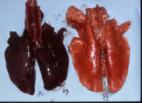
- At post-mortem, the lower parts of the body may be reddened compared to the other parts of the body.
- When the circulation stops after death, the blood tends to gravitate in the blood vessels to the lowest point before the blood has clotted.
- May be difficult to appreciate in most species as many have deeply-pigmented skin and are covered in hair or fur.
- Easily appreciated in the pig.
- Particularly evident the lungs and kidneys once the cadaver has been opened.
- The more dependent lobe or organ is a much deeper red colour than its counterpart on the opposite side.
Post Mortem Imbibition of Blood
- At post-mortem, the surface of organs throughout the body may be diffusely stained by blood.
- Blood pigment tends to diffuse out of the blood cells after death and through the walls of small vessels.
- Taken up by nearby tissues - like blotting paper sucking up ink.
- Can be appreciated on the surface of organs where there is leakage of blood pigment from the serosal or capsular vessels into the nearby tissue.
- Also seen on the surface of other adjacent organs or tissues.
- Post-mortem clots in the large arteries and right ventricle and large arteries undergo autolysis.
- Stains the walls of the vessels and the tissues in contact with the blood vessels.
- Can be quite extensive.
- Obscures any ante-mortem colour changes that may have been present.
- Foetuses that have been dead for some time in utero before their expulsion show diffuse discoloration of all tissues due to imbibition of blood.
- Animals dying from a disease that causes intravascular haemolysis, show very early imbibition staining.
Inbibition of Bile Pigment
- One of the earliest local colour changes.
- Bile salts diffuse out of the gall bladder.
- NOT the same thing as jaundice.
- Generalised discoloration of tissues due to bile pigments seen in the living animal.
Gaseous Distension of the Alimentary Tract
- The intestines and forestomachs may be grossly distended with gas.
- May pop out of the initial incision made in the abdominal wall during PME.
- Caused by the continuation of normal bacterial fermentation in the alimentary tract after death.
- Adjacent organs may show surface pallor.
- Due to the pressure of the distension squeezing blood out of this adjacent tissue.
- If the distension is very great, it may be sufficient to tear the wall of the distended portion, or the diaphragm, allowing herniation of the gut into the thoracic cavity.
- It is possible to distinguish between a post-mortem tear and an ante-mortem rupture by examining the cut ends of the tear or rupture.
- Swelling and haemorrhage will be evident in an ante-mortem rupture due to the host reaction to injury of living tissue.
- Swelling and haemorrhage is absent in a post-mortem tear.
- It is also possible to distinguish tears and ruptures by where the ingesta has come in contact with the parietal or visceral peritoneal surface.
- If this occurred ante-mortem, there will be a reaction to its presence.
- May cling to this surface when attempting to wash it off.
- If this occurred ante-mortem, there will be a reaction to its presence.
- It is possible to distinguish between a post-mortem tear and an ante-mortem rupture by examining the cut ends of the tear or rupture.
- In intestinal distension, the gas tends to rise to the upper part.
- When the coils of intestine are straightened out, the
- Upper portions show distension due to the gas.
- Lower portions show hypostatic congestion.
- This patchy congestion of the intestine should not be confused with an inflammatory condition of the gut.
- When the coils of intestine are straightened out, the
Emphysema
- Invasion by gas producing bacteria.
Autolysis
- Self-digestion of tissue.
- As individual cells die, lysosomal and other enzymes are liberated.
- Digest the tissue, breaking it down.
- Appears similar to necrosis.
- Distinguishable on PME as there is no host inflammatory response to autolysis.
Differences in Tissues
- Particularly marked in the gut and associated glands such as the pancreas.
- These organs are concerned with digestion in life, and their digestive processes continue unregulated after death.
- Autolysis proceeds fairly rapidly in these organs.
- The mucosa of the stomach and intestines may be very soft and come off easily from the submucosa when these organs are opened.
- Other metabolically active tissues also become soft from post-mortem change, e.g. the convoluted tubules of the renal cortex.
- Particularly prominent in PME of the sheep.
- Can be difficult to distinguish this appearance from the toxaemic changes seen in pulpy kidney.
- Also true of other tissues such as the brain and spinal cord.
- Can become almost fluid in texture.
- Serious histopathological deterioration.
- Particularly prominent in PME of the sheep.
- Autolysis proceeds at a more leisurely rate in other tissues such as muscle, connective tissue and skin.
Gross appearance
- Fairly similar to degeneration and necrosis.
- I.e. it is paler than normal.
- Further features include:
- The whole tissue is affected.
- There is no zone of host reaction such as hyperaemia.
- The surface may exude fluid.
- The cut surface tends to be greasy.
- The internal substance tends to be more fluid.
Histological Appearance
- Can be similar to that of necrosis.
Putrefaction
- May be possible to detect putrefaction from outside the animal from the colour and smell.
- Results from the action of putrefactive organisms.
- Most of these are anaerobes.
- Grow best or only in the absence of oxygen.
- Include the Clostridial group of organisms which are found in the large intestine.
- Most of these are anaerobes.
Tissue Degradation
- The body proteins, fats and carbohydrates are attacked by enzymes produced by the putrefactive bacteria.
- Broken down into simpler substances.
- End result being destruction of the carcass (or most of it).
- New substances are produced by the action of putrefactive organisms on tissue protein, carbohydrate and fat, including
- Proteases, polypeptides and amino acids another derived from the protein.
- Indole, skatole and other phenolic compounds.
- Some of these have an unpleasant odour.
- Ammonia is produced from the nitrogen in protein.
- Turns the autolysing muscle undergoing putrefaction , otherwise acid, towards alkalinity.
The Effects of Hydrogen Sulphide
- Hydrogen sulphide is an important product of putrefaction.
- Formed from the breakdown of the sulphur-containing proteins.
- This is responsible for two things noticeable about putrefying tissue.
- Colour
- Smell
Colour
- Putrefaction produces a greenish or blackish discoloration of tissues.
- Colour is due to the development of blackish particles of ferrous sulphide in the tissues.
- The sulphide part is due to the development of hydrogen sulphide in the putrefying tissues.
- The iron part comes from the haemoglobin of the blood.
- Haemoglobin is acted on in putrefaction by bacteria, which split off the iron at the same time as they produce hydrogen sulphide.
- Components combine to form ferrous sulphide.
- Haemoglobin is acted on in putrefaction by bacteria, which split off the iron at the same time as they produce hydrogen sulphide.
- Discolouration therefore depends on the presence of both blood and bacteria.
- To reduce the incidence of discoloration, the animal should be bled out as soon as possible after death.
- Discolouration is obscured in most animals by the covering of hair or fur.
- Except in the pig.
- May still be easily appreciated by parting the fur and noting the green colour on the skin.
- The greenish colour is most noticeable on the skin nearest to the source of putrefactive organisms.
- I.e. since organisms are found in the gut, the abdominal wall is greenest.
- Discolouration resembles in some respects the colour due to melanin (the ordinary pigment of hair and skin).
- Is sometimes called pseudomelanosis.
- Pseudomelanosis occurs not only on the skin on and the large intestine but also on the surface of tissues in contact with it, e.g.
Smell
- Extremely unpleasant
- Smells like rotten eggs.
The Liver
- The liver is very rich in protein and carbohydrates.
- Easily undergoes putrefaction, by:
- Extension of the bacteria across the gut.
- Bacteria growing up the portal vein.
- When bacteria grow up the portal vein, the whole organ is affected rather than just the surface.
- The bacteria produce gas bubbles.
- The whole liver becomes soft, greenish-blackish and foamy in appearance.
- Occurs quite commonly in sheep and cattle, but not often seen in small animals.
- Focal areas of gas formation occur in other tissues undergoing putrefaction.
- The whole liver becomes soft, greenish-blackish and foamy in appearance.
- The bacteria produce gas bubbles.
Other External Features Occuring Post-Mortem
- Occur post-mortem or very shortly before death in very weak animals.
- Whitish fly larvae in the mouth.
- Look like rice grains.
- Deposited after death and appear within hours.
- Removal of tissue by rats or crows.
- The tip of a protruding tongue may be bluish in colour and dry from the clenched jaws.
Agonal Changes
- Occur around the time of death/ time of irreversible circulatory failure.
- Often leads to vascular congestion e.g. in the lungs.
- May be exacerbated by hypostatic changes, causing pooling of blood in dependent sites.
- Kidneys, liver and pancreas may similarly be affected by vascular congestion.
- The spleen is particularly susceptible to extreme congestion relating to barbiturate euthanasia.
- Also seen in animals dying under anaesthesia.
- Crystal deposition on the endocardium is another common barbiturate associated change.
- Agonal regurgitation of GI contents can also occur resulting in food material being present in the airways and possibly alveoli.
