Difference between revisions of "Macrophages"
Fiorecastro (talk | contribs) |
Kate English (talk | contribs) |
||
| (23 intermediate revisions by 3 users not shown) | |||
| Line 1: | Line 1: | ||
| + | [[Image:LH Macrophage Histology.jpg|thumb|right|125px|<p>'''Macrophage'''</p><sup>© Nottingham Uni</sup>]] | ||
==Introduction== | ==Introduction== | ||
| − | + | Macrophages are large, round cells that contain a central round nucleus and have abundant clear, often vacuolated, cytoplasm. Macrophages acts as sentinel cells; they have a role in destroying bacteria, protozoa and tumour cells, and release substances that act upon other immune cells. Macrophages are phagocytic, long lived and are found throughout the body. | |
| − | Macrophages are large, round cells that contain a central round nucleus and have abundant clear, often vacuolated, cytoplasm. Macrophages acts as sentinel cells; they have a role in destroying | ||
==Development== | ==Development== | ||
Macrophages are either derived from blood borne [[Monocytes|monocytes]] which have migrated into tissue and differentiated, or from dividing macrophages within the tissue. | Macrophages are either derived from blood borne [[Monocytes|monocytes]] which have migrated into tissue and differentiated, or from dividing macrophages within the tissue. | ||
| − | + | ===Locations=== | |
| − | ==Locations== | ||
<p>Macrophages are present throughout the body with large numbers in the [[Lymph Nodes - Anatomy & Physiology|lymph nodes]], [[Bone Marrow - Anatomy & Physiology|bone marrow]] and [[Spleen - Anatomy & Physiology|spleen]]. In connective tissue macrophages are fixed and referred to as tissue histocytes. Sentinel macrophages in the lung are called alveolar macrophages, while in the [[Liver - Anatomy & Physiology|liver]] they are called Kupffer cells. In the brain they are microglia with long cytoplasmic processes.</p> | <p>Macrophages are present throughout the body with large numbers in the [[Lymph Nodes - Anatomy & Physiology|lymph nodes]], [[Bone Marrow - Anatomy & Physiology|bone marrow]] and [[Spleen - Anatomy & Physiology|spleen]]. In connective tissue macrophages are fixed and referred to as tissue histocytes. Sentinel macrophages in the lung are called alveolar macrophages, while in the [[Liver - Anatomy & Physiology|liver]] they are called Kupffer cells. In the brain they are microglia with long cytoplasmic processes.</p> | ||
| − | ==Variations== | + | ===Variations=== |
| − | ===Epithelioid cells=== | + | ====Epithelioid cells==== |
| − | + | * Look like squamous epithelial cells with a pink (eosinophilic) cytoplasm and indistinct borders. | |
| + | * May be binucleate. | ||
| + | * Tend to remain in the lesion. | ||
| + | * It is thought by some people that they may not phagocytose, but instead secrete substances directed against the foreign agent. | ||
| − | ===Giant cells=== | + | ====Giant cells==== |
| − | <p>Macrophages can fuse together to form giant cells (Langhan’s cells), which with their greater cytoplasmic volume and number of lysosomes are able to engulf and deal with large foreign particles/bodies. They are thought to form when two or more macrophages attempt to engulf the same organism; the resulting cell can contain between two to several hundred nuclei per cell).</p><p>The nuclei can be scattered throughout the cytoplasm, clumped in the centre in foreign body granulomas or appear in a horseshoe shape at the periphery of the cytoplasm at one end in | + | <p>Macrophages can fuse together to form giant cells (Langhan’s cells), which with their greater cytoplasmic volume and number of lysosomes are able to engulf and deal with large foreign particles/bodies. They are thought to form when two or more macrophages attempt to engulf the same organism; the resulting cell can contain between two to several hundred nuclei per cell).</p><p>The nuclei can be scattered throughout the cytoplasm, clumped in the centre in foreign body granulomas or appear in a horseshoe shape at the periphery of the cytoplasm at one end in tuberculosis and some other granulomas. In the past, the morphology of these giant cells was correlated with the agent responsible for inflammation, although the distinction is not absolute.</p> |
==Actions== | ==Actions== | ||
| − | + | <p>Macrophages are phagocytic and take up particles and cell debris by endocytosis, as well has engulfing pathogens like bacteria. These are then present in the macrophage inside phagosomes. Lysosomes present in the cytoplasm then bind with the phagosome and release their contents which degrade/digest its contents.</p> | |
| − | + | <p>Macrophages also act as '''antigen presenting cells''' taking antigens to lymph nodes to present to T cells. MHC II ([[Major Histocompatability Complexes|major histocompatibility complex]] II) proteins on their surface allow them to interact with helper T cells (CD4). Short peptide segments from foreign cells are presented with MHC II which activates the [[T cells|T cell]].</p> | |
| − | <p>Macrophages are | + | <p>To migrate through connective tissue they release proteases and glycoaminoglycanases.</p> |
| − | |||
| − | |||
| − | |||
| − | |||
| − | |||
| − | |||
| − | |||
| − | |||
| − | |||
| − | |||
| − | <p>Macrophages also act as '''antigen presenting cells''' taking antigens to lymph nodes to present to T cells. MHC II ([[Major Histocompatability Complexes|major histocompatibility complex]] II) proteins on their surface allow them to interact with helper T cells (CD4). Short peptide segments from foreign cells are presented with MHC II which activates the [[T cells|T cell]] | ||
| − | |||
| − | |||
| − | |||
| − | |||
| − | |||
| − | |||
| − | |||
| − | |||
| − | <p>To migrate through connective tissue they release proteases and glycoaminoglycanases | ||
| − | |||
| − | |||
===Role in pathology=== | ===Role in pathology=== | ||
| − | <p>Macrophages are seen in both [[Acute Inflammation|acute]] and [[Chronic Inflammation| chronic inflammation]].</p> If large numbers of macrophages are found in chronic inflammatory processes, it implies the inability to eliminate the causal organism e.g. [[:Category:Mycobacterium species| | + | <p>Macrophages are seen in both [[Acute Inflammation|acute]] and [[Chronic Inflammation| chronic inflammation]].</p> If large numbers of macrophages are found in chronic inflammatory processes, it implies the inability to eliminate the causal organism e.g. [[:Category:Mycobacterium species|Mycobacterium (TB)]], [[:Category:Actinobacillus species|Actinobacillus]], fungi, parasites and foreign bodies. |
<br> | <br> | ||
| − | + | [[Category:Blood_Cells]] [[Category:Kate English reviewing]] | |
| − | |||
| − | |||
| − | |||
| − | |||
| − | |||
| − | |||
| − | |||
| − | |||
| − | |||
| − | [[Category:Blood_Cells]] [[Category: | ||
Revision as of 12:52, 16 February 2011
Introduction
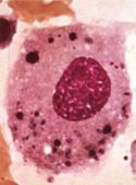
Macrophages are large, round cells that contain a central round nucleus and have abundant clear, often vacuolated, cytoplasm. Macrophages acts as sentinel cells; they have a role in destroying bacteria, protozoa and tumour cells, and release substances that act upon other immune cells. Macrophages are phagocytic, long lived and are found throughout the body.
Development
Macrophages are either derived from blood borne monocytes which have migrated into tissue and differentiated, or from dividing macrophages within the tissue.
Locations
Macrophages are present throughout the body with large numbers in the lymph nodes, bone marrow and spleen. In connective tissue macrophages are fixed and referred to as tissue histocytes. Sentinel macrophages in the lung are called alveolar macrophages, while in the liver they are called Kupffer cells. In the brain they are microglia with long cytoplasmic processes.
Variations
Epithelioid cells
- Look like squamous epithelial cells with a pink (eosinophilic) cytoplasm and indistinct borders.
- May be binucleate.
- Tend to remain in the lesion.
- It is thought by some people that they may not phagocytose, but instead secrete substances directed against the foreign agent.
Giant cells
Macrophages can fuse together to form giant cells (Langhan’s cells), which with their greater cytoplasmic volume and number of lysosomes are able to engulf and deal with large foreign particles/bodies. They are thought to form when two or more macrophages attempt to engulf the same organism; the resulting cell can contain between two to several hundred nuclei per cell).
The nuclei can be scattered throughout the cytoplasm, clumped in the centre in foreign body granulomas or appear in a horseshoe shape at the periphery of the cytoplasm at one end in tuberculosis and some other granulomas. In the past, the morphology of these giant cells was correlated with the agent responsible for inflammation, although the distinction is not absolute.
Actions
Macrophages are phagocytic and take up particles and cell debris by endocytosis, as well has engulfing pathogens like bacteria. These are then present in the macrophage inside phagosomes. Lysosomes present in the cytoplasm then bind with the phagosome and release their contents which degrade/digest its contents.
Macrophages also act as antigen presenting cells taking antigens to lymph nodes to present to T cells. MHC II (major histocompatibility complex II) proteins on their surface allow them to interact with helper T cells (CD4). Short peptide segments from foreign cells are presented with MHC II which activates the T cell.
To migrate through connective tissue they release proteases and glycoaminoglycanases.
Role in pathology
Macrophages are seen in both acute and chronic inflammation.
If large numbers of macrophages are found in chronic inflammatory processes, it implies the inability to eliminate the causal organism e.g. Mycobacterium (TB), Actinobacillus, fungi, parasites and foreign bodies.
