Difference between revisions of "Osteochondrosis"
| (6 intermediate revisions by 3 users not shown) | |||
| Line 1: | Line 1: | ||
| − | [[Image:Pig elbow osteochondrosis.jpg|right|thumb| | + | {{OpenPagesTop}} |
| − | [[Image:Osteochondrosis dissecans.jpg|right|thumb| | + | ==Introduction== |
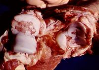
| + | [[Image:Pig elbow osteochondrosis.jpg|right|thumb|200px|<small><center>Osteochondrosis in pig elbow (Image sourced from Bristol Biomed Image Archive with permission)</center></small>]] | ||
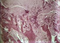
| + | [[Image:Osteochondrosis dissecans.jpg|right|thumb|200px|<small><center>Osteochondrosis dissecans (Image sourced from Bristol Biomed Image Archive with permission)</center></small>]] | ||
| + | Osteochondrosis describes a defect in [[Cartilage - Anatomy & Physiology|endochondral ossification]] of the epiphyseal cartilage. The term is currently used to describe the clinical manifestation of the disorder. The term '''dyschondrodysplasia''' is preferred when referring to early lesions because primary lesions are seen in the cartilage. | ||
| − | + | It is one of the '''most prevalent and important developmental orthopaedic diseases''' of dogs and horses, and is seen in growing animals. '''Large breeds of dogs''' present at 4-8 months, '''horses''' from weeks to 2 years, and '''pigs''' from 5-7 months. | |
| − | |||
| − | |||
| − | |||
| − | |||
| − | |||
| − | |||
| − | |||
| − | |||
| − | |||
| − | |||
| − | |||
| − | |||
| − | + | It involves both the [[Bones - Anatomy & Physiology|growth plate]] and the immature [[Joints - Anatomy & Physiology#Articular cartilage|joint cartilage]]. Lesions are '''bilateral in 70% of cases''' but lameness is often unilateral. | |
| − | + | The condition mainly affects '''articular growth cartilage''', but the metaphysis may also be involved. If the physeal metaphyseal cartilage is affected, bone contours and longitudinal growth are disturbed. Involvement of articular cartilage at the periphery of joint surfaces leads to regressive changes at the joint margins, '''dissecting lesions''', and the '''formation of flaps'''. Central articular lesions, because of weight-bearing effects, involve focal retention of cartilage within the subchondral bone ('''Subchondral Bone Cysts'''). Axial skeletal involvement includes vertebral articular facets, and this may lead to '''stenosis of the vertebral canal''' and, ultimately, ataxia and proprioceptive deficits (ie, [[Cervical Vertebral Stenotic Myelopathy|wobbler syndrome]] in horses). | |
| − | |||
| − | |||
| − | |||
| − | |||
| − | |||
| − | |||
| − | |||
| − | |||
| − | |||
| − | |||
| − | |||
| − | |||
| − | |||
| − | |||
| − | |||
| − | |||
| − | |||
| − | |||
| − | |||
| − | |||
| − | |||
| − | |||
| − | |||
| − | |||
| − | |||
| − | |||
| − | |||
| − | |||
| − | |||
| − | + | The exact cause is unknown, but contributing factors include: | |
| − | + | '''excessive nutrition, rapid growth, trauma, mineral imbalance and a hereditary component'''. | |
| − | |||
| − | |||
| − | |||
| + | In dogs, '''[[Elbow Dysplasia|ununited anconeal process and fragmented coronoid process]]''' may be related conditions, linked to separation of the epiphysis from the metaphysis likely due to osteochondrosis which predisposes the growth plate to failure following trauma. | ||
| + | ==Osteochondrosis dissecans (OCD)== | ||
| + | This is one manifestation of osteochondrosis, and involves '''retention of cartilage cores''' at the articular surface. | ||
| + | |||
| + | White, wedge-shaped areas of [[Retained Cartilage Cores|retained cartilage]] are found in the metaphysis. Clefts lead to the separation of cartilage from bone and the formation of '''flaps or free joint mice''' which may interfere with joint function. | ||
| + | |||
| + | This condition may lead to [[Angular Limb Deformity|angular limb deformities]] and [[Degenerative Joint Disease|degenerative joint disease]] and may be present together with '''synovitis'''. | ||
| + | |||
| + | Predilection sites include: | ||
| + | |||
| + | <u>In dogs:</u> proximal humerus, lateral femoral condyle and the coronoid process of ulna | ||
| + | |||
| + | <u>In pigs:</u> humeral and medial femoral condyles and anconeal process of elbow | ||
| + | |||
| + | <u>In horses:</u> femoropatellar joint, tibiotarsal joint, fetlock joint and the shoulder. | ||
| + | |||
| + | ==Clinical Signs== | ||
| + | Clinical signs are difficult to characterise due to the variety of sites and lesions involved. | ||
| + | |||
| + | In '''horses''', signs may begin with '''mild stiffness and lameness''' progressing to pain and loss of performance. The most common sign is '''non-painful distention of the affected joints'''. | ||
| + | |||
| + | '''Foals''' tend to spend more time lying down and show stiffness, an upright limb conformation and a difficulty in keeping up with other animals in the paddock. Signs in older horses are usually linked to the '''onset of training''' and include stiffness, flexion responses and variable lameness. Marked lameness is not usually a feature. | ||
| + | |||
| + | Shoulder OC cases usually show more severe lameness and possibly some muscle atrophy. | ||
| + | |||
| + | '''Dogs''' show lameness, joint effusion and a reduced range of motion in the affected joints. | ||
| + | |||
| + | ==Diagnosis== | ||
| + | Clinical diagnosis is usually made on the basis of clinical signs and signalment. | ||
| + | |||
| + | '''Radiographic examination''' is usually useful in making a diagnosis, although early lesions involving cartilage without significant subchondral bone damage will not be visualised. Oblique views may be helpful. | ||
| + | Radiographic signs include: '''flattening of joint surfaces, subchondral bone lucency or sclerosis, osteophytosis, joint effusion, and joint mice'''. | ||
| + | |||
| + | '''Contrast radiography''' can be used to delineate cartilage flaps. '''Ultrasonography''' may delineate articular damage and joint mice. | ||
| + | |||
| + | '''Arthroscopy''' is the most accurate way to confirm the diagnosis and most sites are accessible. | ||
| + | |||
| + | Evaluation of '''synovial fluid''' will help rule out inflammatory causes of swollen joints. | ||
| + | |||
| + | '''Histological findings''' include: lesion filled with granulation tissue (fibroplasia), surrounding thickened bone spicules, cap of thickened articular cartilage over the defect. | ||
| + | |||
| + | ==Treatment== | ||
| + | Treatment depends on the site and severity of signs. | ||
| + | |||
| + | '''In horses''', mild cases resolve spontaneously and a '''conservative approach''' may be adequate. Young animals should have their '''exercise restricted''' and their '''feed intake reduced''' to slow growth. Correcting the diet may not aid resolution but may help prevent further cases on a stud farm. '''Intra-articular medication''' with hyaluronic acid may be beneficial, and injection of long-acting corticosteroids may help reduce swelling and improve any associated synovitis. | ||
| + | |||
| + | '''Surgical cases''' can be treated '''arthroscopically'''. Damaged cartilage and joint mice are removed and the bone overlying the lesion is curetted and the joint flushed extensively. | ||
| + | |||
| + | '''Prognosis is good''' except in cases with severe joint disruption and secondary osteoarthritis. | ||
| + | |||
| + | '''Shoulder osteochondrosis''' is more problematic and the prognosis is guarded. | ||
| + | |||
| + | '''In dogs''', treatment involves '''surgical removal of flaps and joint mice''' and curettage of subchondral bone to stimulate fibrocartilage formation. The joint can also be management '''conservatively''' and the animal given [[NSAIDs]] at times of need. Joint fluid modifiers such as polysulfated glycosaminoglycans may also help prevent cartilage degeneration. | ||
| + | |||
| + | '''Prognosis''' for recovery is excellent for the shoulders, good for the stifle joint, and fair for the elbow and tarsal joints. Concomitant signs of degenerative joint disease, other joint conditions, or instability deleteriously affect recovery. | ||
| + | |||
| + | {{Learning | ||
| + | |flashcards = [[Equine Orthopaedics and Rheumatology Q&A 14]] | ||
| + | }} | ||
| + | |||
| + | ==References== | ||
| + | Kahn, C. (2005) '''Merck veterinary manual''' ''Merck and co'' | ||
| + | |||
| + | Morgan, J. (2000) '''Hereditary bone and joint diseases in the dog''' ''Schültersche'' | ||
| + | |||
| + | |||
| + | {{review}} | ||
| + | |||
| + | {{OpenPages}} | ||
| + | |||
| + | [[Category:Musculoskeletal Diseases - Pig]] | ||
| + | [[Category:Expert Review]] | ||
[[Category:Joints - Developmental Pathology]] | [[Category:Joints - Developmental Pathology]] | ||
| + | [[Category:Musculoskeletal Diseases - Dog]] | ||
| + | [[Category:Musculoskeletal Diseases - Horse]] | ||
Latest revision as of 16:20, 18 July 2012
Introduction
Osteochondrosis describes a defect in endochondral ossification of the epiphyseal cartilage. The term is currently used to describe the clinical manifestation of the disorder. The term dyschondrodysplasia is preferred when referring to early lesions because primary lesions are seen in the cartilage.
It is one of the most prevalent and important developmental orthopaedic diseases of dogs and horses, and is seen in growing animals. Large breeds of dogs present at 4-8 months, horses from weeks to 2 years, and pigs from 5-7 months.
It involves both the growth plate and the immature joint cartilage. Lesions are bilateral in 70% of cases but lameness is often unilateral.
The condition mainly affects articular growth cartilage, but the metaphysis may also be involved. If the physeal metaphyseal cartilage is affected, bone contours and longitudinal growth are disturbed. Involvement of articular cartilage at the periphery of joint surfaces leads to regressive changes at the joint margins, dissecting lesions, and the formation of flaps. Central articular lesions, because of weight-bearing effects, involve focal retention of cartilage within the subchondral bone (Subchondral Bone Cysts). Axial skeletal involvement includes vertebral articular facets, and this may lead to stenosis of the vertebral canal and, ultimately, ataxia and proprioceptive deficits (ie, wobbler syndrome in horses).
The exact cause is unknown, but contributing factors include: excessive nutrition, rapid growth, trauma, mineral imbalance and a hereditary component.
In dogs, ununited anconeal process and fragmented coronoid process may be related conditions, linked to separation of the epiphysis from the metaphysis likely due to osteochondrosis which predisposes the growth plate to failure following trauma.
Osteochondrosis dissecans (OCD)
This is one manifestation of osteochondrosis, and involves retention of cartilage cores at the articular surface.
White, wedge-shaped areas of retained cartilage are found in the metaphysis. Clefts lead to the separation of cartilage from bone and the formation of flaps or free joint mice which may interfere with joint function.
This condition may lead to angular limb deformities and degenerative joint disease and may be present together with synovitis.
Predilection sites include:
In dogs: proximal humerus, lateral femoral condyle and the coronoid process of ulna
In pigs: humeral and medial femoral condyles and anconeal process of elbow
In horses: femoropatellar joint, tibiotarsal joint, fetlock joint and the shoulder.
Clinical Signs
Clinical signs are difficult to characterise due to the variety of sites and lesions involved.
In horses, signs may begin with mild stiffness and lameness progressing to pain and loss of performance. The most common sign is non-painful distention of the affected joints.
Foals tend to spend more time lying down and show stiffness, an upright limb conformation and a difficulty in keeping up with other animals in the paddock. Signs in older horses are usually linked to the onset of training and include stiffness, flexion responses and variable lameness. Marked lameness is not usually a feature.
Shoulder OC cases usually show more severe lameness and possibly some muscle atrophy.
Dogs show lameness, joint effusion and a reduced range of motion in the affected joints.
Diagnosis
Clinical diagnosis is usually made on the basis of clinical signs and signalment.
Radiographic examination is usually useful in making a diagnosis, although early lesions involving cartilage without significant subchondral bone damage will not be visualised. Oblique views may be helpful. Radiographic signs include: flattening of joint surfaces, subchondral bone lucency or sclerosis, osteophytosis, joint effusion, and joint mice.
Contrast radiography can be used to delineate cartilage flaps. Ultrasonography may delineate articular damage and joint mice.
Arthroscopy is the most accurate way to confirm the diagnosis and most sites are accessible.
Evaluation of synovial fluid will help rule out inflammatory causes of swollen joints.
Histological findings include: lesion filled with granulation tissue (fibroplasia), surrounding thickened bone spicules, cap of thickened articular cartilage over the defect.
Treatment
Treatment depends on the site and severity of signs.
In horses, mild cases resolve spontaneously and a conservative approach may be adequate. Young animals should have their exercise restricted and their feed intake reduced to slow growth. Correcting the diet may not aid resolution but may help prevent further cases on a stud farm. Intra-articular medication with hyaluronic acid may be beneficial, and injection of long-acting corticosteroids may help reduce swelling and improve any associated synovitis.
Surgical cases can be treated arthroscopically. Damaged cartilage and joint mice are removed and the bone overlying the lesion is curetted and the joint flushed extensively.
Prognosis is good except in cases with severe joint disruption and secondary osteoarthritis.
Shoulder osteochondrosis is more problematic and the prognosis is guarded.
In dogs, treatment involves surgical removal of flaps and joint mice and curettage of subchondral bone to stimulate fibrocartilage formation. The joint can also be management conservatively and the animal given NSAIDs at times of need. Joint fluid modifiers such as polysulfated glycosaminoglycans may also help prevent cartilage degeneration.
Prognosis for recovery is excellent for the shoulders, good for the stifle joint, and fair for the elbow and tarsal joints. Concomitant signs of degenerative joint disease, other joint conditions, or instability deleteriously affect recovery.
| Osteochondrosis Learning Resources | |
|---|---|
 Test your knowledge using flashcard type questions |
Equine Orthopaedics and Rheumatology Q&A 14 |
References
Kahn, C. (2005) Merck veterinary manual Merck and co
Morgan, J. (2000) Hereditary bone and joint diseases in the dog Schültersche
| This article has been peer reviewed but is awaiting expert review. If you would like to help with this, please see more information about expert reviewing. |
Error in widget FBRecommend: unable to write file /var/www/wikivet.net/extensions/Widgets/compiled_templates/wrt6746ca47a8f004_25767397 Error in widget google+: unable to write file /var/www/wikivet.net/extensions/Widgets/compiled_templates/wrt6746ca47b1acd9_16296282 Error in widget TwitterTweet: unable to write file /var/www/wikivet.net/extensions/Widgets/compiled_templates/wrt6746ca47c17771_67802731
|
| WikiVet® Introduction - Help WikiVet - Report a Problem |

