Difference between revisions of "Colon - Anatomy & Physiology"
Fiorecastro (talk | contribs) |
Thepollycat (talk | contribs) m (→Links) |
||
| (2 intermediate revisions by 2 users not shown) | |||
| Line 22: | Line 22: | ||
===Equine=== | ===Equine=== | ||
| − | For information on the equine colon, see: [[Alimentary System - | + | For information on the equine colon, see: [[Equine Alimentary System - Anatomy & Physiology|equine alimentary system]]. |
===Porcine=== | ===Porcine=== | ||
| Line 39: | Line 39: | ||
{{Template:Learning | {{Template:Learning | ||
|flashcards = [[Colon_- Anatomy & Physiology_-_Flashcards|Colon Flashcards]] | |flashcards = [[Colon_- Anatomy & Physiology_-_Flashcards|Colon Flashcards]] | ||
| − | |videos = [ | + | |videos = [http://stream2.rvc.ac.uk/Anatomy/bovine/Pot0048.mp4 The Small and Large intestine of the Ruminant]<br>[http://stream2.rvc.ac.uk/Frean/Pony/left_topography.mp4 Left Sided topography of the Equine abdomen]<br>[http://stream2.rvc.ac.uk/Frean/Pony/right_topography.mp4 Right sided topography of the Equine Abdomen]<br>[http://stream2.rvc.ac.uk/Anatomy/feline/pot0357.mp4 The Feline Abdomen]<br>[http://stream2.rvc.ac.uk/Frean/sheep/LargeSmallIntestine.mp4 Small and Large intestine of the Sheep]<br>[http://stream2.rvc.ac.uk/Frean/sheep/RightSideTopography.mp4 Right sided topography of the Ovine Abdomen]<br>[[Video:Porcine abdomen dissection|Dissection of the porcine abdomen]] |
| − | |||
|powerpoints = [[Gastrointestinal Tract Histology resource|Histology of the colon - see part 1]] | |powerpoints = [[Gastrointestinal Tract Histology resource|Histology of the colon - see part 1]] | ||
}} | }} | ||
| − | + | {{OpenPages}} | |
| − | |||
| − | |||
[[Category:Large Intestine - Anatomy & Physiology]] | [[Category:Large Intestine - Anatomy & Physiology]] | ||
[[Category:A&P Done]] | [[Category:A&P Done]] | ||
Revision as of 13:44, 13 August 2012
Overview
The colon is a site of microbial fermentation, the relative importance of this is species dependent. The colon can be divided into the following portions; Ascending, transverse and descending.
Structure
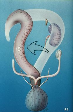
The following anatomical arrangement is found only in cats and dogs, see species differences. The ascending colon continues from the iluem at the ileocolic junction. It runs to the right of the cranial mesenteric artery in a caudal to cranial direction. At the cranial border of the mesentery it turns medially to become the transverse colon. The transverse colon runs from the right side of the abdomen to the left side of the abdomen. Cranial to the transverse colon is the stomach, and caudal to it is the small intestine and cranial mesenteric artery. The descending colon continues on from the transverse colon running caudally on the left. It then passes more medially as it enters the pelvic cavity. Upon entering the pelvic cavity it is continued as the rectum.
Function
The colon is a site of microbial fermentation and absorption of the products of microbial fermentation, volatile fatty acids(VFAs). Transportation is also important here. Motility in most species is brought about by segmentation and peristalsis. Antiperistalsis also occurs and is particularly important in horses, ruminants and rodents. Chyme is transported towards the small intestine so as to fill the caecum. In the horse, the antiperistaltic movements delay the movement of chyme from the ventral to the dorsal colon, which increases the time chyme is available for fermentation in the ventral colon. Mass movements move the content of the large intestine into the rectum.
Species Differences
Ruminant
The ascending colon is the longest part of the colon and is composed of three parts. The ansa proximalis has a sigmoid flexure that passes around the caudal border of the mesentry to the left side of the root of the mesentry. The ansa spiralis consists of two centripetal turns and two centrifugal turns in the ox. There are three turns in the sheep and four in the goat. In the ox, the ansa spiralis is a flat disc, whilst in the small ruminants it takes the form of a cone. The ansa distalis goes back around the caudal border of the mesentry, to the right side of the root of mesentry. It then passes cranially adjacent to the mesentery until it reaches the cranial border of the mesentery. The transverse colon crosses the midline of the abdomen, from right to left at the cranial border of the mesentery. The descending colon continues caudally to the rectum and anus. It has a sigmoid flexure before it enters the pelvic cavity.
Development The ox's ascending colon expands caudally around the root of the mesentery on the left side of the mesentery (compare to horse, where it expands cranially).
Equine
For information on the equine colon, see: equine alimentary system.
Porcine
The arrangement of the transverse and descending colon is similar to that of the dog and cat, but the ascending colon is different. The ascending colon is elongated and coiled to form a cone-shaped organ. The base of the cone is attached to the dorsal abdominal wall and the apex points ventrally. The position of the ascending colon varies with filling of the stomach. From the caecum, there are clockwise centripetal turns to the apex of the cone. Then the centrifugal turns run anti-clockwise on the inside of the cone. Centripetal turns have two taenia, whilst centrifugal turns have none.
Histology
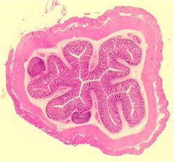
The mucosa of the colon is thick and has long glands with columnar epithelium. The Submucosa has large lymphatic nodules which may interrupt the lamina muscularis. The lamina muscularis is incomplete in the colon.
Links
Click here for information on the pathology of the Small and Large Intestines
| Colon - Anatomy & Physiology Learning Resources | |
|---|---|
 Test your knowledge using flashcard type questions |
Colon Flashcards |
 Selection of relevant videos |
The Small and Large intestine of the Ruminant Left Sided topography of the Equine abdomen Right sided topography of the Equine Abdomen The Feline Abdomen Small and Large intestine of the Sheep Right sided topography of the Ovine Abdomen Dissection of the porcine abdomen |
 Selection of relevant PowerPoint tutorials |
Histology of the colon - see part 1 |
Error in widget FBRecommend: unable to write file /var/www/wikivet.net/extensions/Widgets/compiled_templates/wrt69a0cce39aece7_01648838 Error in widget google+: unable to write file /var/www/wikivet.net/extensions/Widgets/compiled_templates/wrt69a0cce3a06657_51528643 Error in widget TwitterTweet: unable to write file /var/www/wikivet.net/extensions/Widgets/compiled_templates/wrt69a0cce3a53db9_11658412
|
| WikiVet® Introduction - Help WikiVet - Report a Problem |

