Difference between revisions of "Integument of Exotic Species - Anatomy & Physiology"
Fiorecastro (talk | contribs) |
(New page: {{toplink |linkpage =Integumentary - Anatomy & Physiology |linktext =INTEGUMENTARY SYSTEM |maplink = Integumentary System (Content Map) - Anatomy & Physiology |pagetype =Anatomy }} <br> ==...) |
||
| (9 intermediate revisions by 3 users not shown) | |||
| Line 1: | Line 1: | ||
| − | {{ | + | {{toplink |
| + | |linkpage =Integumentary - Anatomy & Physiology | ||
| + | |linktext =INTEGUMENTARY SYSTEM | ||
| + | |maplink = Integumentary System (Content Map) - Anatomy & Physiology | ||
| + | |pagetype =Anatomy | ||
| + | }} | ||
| + | <br> | ||
==Birds== | ==Birds== | ||
| Line 64: | Line 70: | ||
The '''cere''' is situated at the base of the upper beak and is composed of keratinised skin. The colour of the cere is influenced by diet and hormones. | The '''cere''' is situated at the base of the upper beak and is composed of keratinised skin. The colour of the cere is influenced by diet and hormones. | ||
| − | The beak has two holes called '''nares''' (nostrils) which connect to the hollow inner beak and thence to the [[Cardiorespiratory System | + | The beak has two holes called '''nares''' (nostrils) which connect to the hollow inner beak and thence to the [[Cardiorespiratory System - Anatomy & Physiology|respiratory system]]. The nares are usually located on the dorsal beak. In some birds, they are located at the base of the beak in the '''cere'''. |
In some species of bird, the tip of the beak is hard, dead tissue used for heavy-duty tasks such as cracking nuts or killing prey. In other species of bird, such as ducks, the tip of the bill is sensitive and contains nerves, for locating things by touch. The beak is worn down by use, so it grows continuously throughout the bird's life. | In some species of bird, the tip of the beak is hard, dead tissue used for heavy-duty tasks such as cracking nuts or killing prey. In other species of bird, such as ducks, the tip of the bill is sensitive and contains nerves, for locating things by touch. The beak is worn down by use, so it grows continuously throughout the bird's life. | ||
| Line 160: | Line 166: | ||
The skin consists of [[Skin - Anatomy & Physiology#Epidermis|epidermal]] cells and '''scales''' (in most species) which are covered by a protective outer mucus '''cuticle'''. Most fish are covered in a protective layer of slime (mucus). | The skin consists of [[Skin - Anatomy & Physiology#Epidermis|epidermal]] cells and '''scales''' (in most species) which are covered by a protective outer mucus '''cuticle'''. Most fish are covered in a protective layer of slime (mucus). | ||
| − | [[image: Fish scales.jpg|thumb|250px| | + | [[image: Fish scales.jpg|thumb|250px|left|Scales of Bitterling (''Rhodeus amarus'')from the hindflank. The bluegreen colour of the tailstripe comes from the epidermis, background colour from the dermis. Photograph by Piet Spaans]] |
There are four types of fish scales. | There are four types of fish scales. | ||
| Line 167: | Line 173: | ||
# '''Cycloid scales''' are small oval-shaped scales with growth rings. Bowfin and remora have cycloid scales. | # '''Cycloid scales''' are small oval-shaped scales with growth rings. Bowfin and remora have cycloid scales. | ||
# '''Ctenoid scales''' are similar to the cycloid scales, with growth rings. They are distinguished by spines that cover one edge. Halibut have this type of scale. | # '''Ctenoid scales''' are similar to the cycloid scales, with growth rings. They are distinguished by spines that cover one edge. Halibut have this type of scale. | ||
| + | |||
| + | |||
| + | |||
| + | |||
| + | |||
| + | |||
| + | |||
| + | |||
| + | |||
| + | |||
| Line 190: | Line 206: | ||
Unlike mammalian epidermal cells, fish epidermal cells at all levels are capable of cell division. During wound healing, cells migrate rapidly to cover any defect and restore the waterproof integrity of the skin. | Unlike mammalian epidermal cells, fish epidermal cells at all levels are capable of cell division. During wound healing, cells migrate rapidly to cover any defect and restore the waterproof integrity of the skin. | ||
| − | + | ||
====Dermis==== | ====Dermis==== | ||
The dermis of fish consists of a '''''stratum spongiosum''''' and a deeper '''''stratum compactum'''''. It also contains '''chromatophores, mechanoreceptors, chemoreceptors''' and '''electroreceptors'''. | The dermis of fish consists of a '''''stratum spongiosum''''' and a deeper '''''stratum compactum'''''. It also contains '''chromatophores, mechanoreceptors, chemoreceptors''' and '''electroreceptors'''. | ||
| Line 200: | Line 216: | ||
*In fish, the '''lateral line''' is a sense organ used to detect movement and vibration in the surrounding water. Lateral lines are usually visible as faint lines running lengthways down each side, from the vicinity of the gill covers to the base of the tail. Sometimes parts of the lateral organ are modified into electroreceptors, which are organs used to detect electrical impulses. It is possible that vertebrates such as sharks use the lateral organs to detect magnetic fields as well. Most amphibian larvae and some adult amphibians also have a '''lateral organ'''. | *In fish, the '''lateral line''' is a sense organ used to detect movement and vibration in the surrounding water. Lateral lines are usually visible as faint lines running lengthways down each side, from the vicinity of the gill covers to the base of the tail. Sometimes parts of the lateral organ are modified into electroreceptors, which are organs used to detect electrical impulses. It is possible that vertebrates such as sharks use the lateral organs to detect magnetic fields as well. Most amphibian larvae and some adult amphibians also have a '''lateral organ'''. | ||
| − | + | [[image: Dorsal fin.jpg|thumb|250px|right|Dorsal fin of a Chubb (''Leuciscus cephalus''). Photograph by Tino Strauss]] | |
*'''Fins''' are modified structures of the skin that aid locomotion and balance in the water (and on land). | *'''Fins''' are modified structures of the skin that aid locomotion and balance in the water (and on land). | ||
*'''Barbels''' are specialised sense organs around the mouth which contain many chemoreceptors | *'''Barbels''' are specialised sense organs around the mouth which contain many chemoreceptors | ||
| − | |||
| − | |||
| − | |||
| − | |||
| − | |||
| − | |||
| − | |||
| − | |||
| − | |||
| − | |||
| − | |||
Revision as of 21:37, 13 August 2009
|
|
Birds
Avian Skin
The general make-up of the avian skin is similar to that of mammals, having an epidermis a dermis and a subcutaneous layer. In comparison, however, it is much thinner, effectively glandless and contains feathers. Generally, the skin is thin enough to be transparent, aiding examination of superficial internal organs including the liver. The reduced thickness of the skin is an adaptation to flying, minimising the weight of the bird.
Epidermis
The epidermis consists of 3 layers:
- The basal (germinative) layer stratum basale
- Intermediate layer
- Superficial (cornified) layer - stratum corneum
Striated muscles in the epidermis move the skin. The epidermis secretes a thin lipid film that helps to maintain the plumage, mimicking a holocrine sebaceous gland, a feature unique to birds.
Dermis
The dermis is divided into:
- A superficial layer which varies in thickness depending on position on the body and age of the bird. This layer contains loosely arranged layers of collagen in interwoven bundles.
- A deep layer containing fat, feather follicles, smooth muscles that control the movement of the feathers, blood vessels and nerves that supply the dermis and epidermis.
The subcutaneous layer is formed mainly by loose connective tissue. It also contains fat, both as a layer, and in discrete fat bodies. These are readily observed as yellow deposits beneath the skin.
Areas of fat deposition vary from species to species (high in aquatic birds) and the time of year (pre-migration deposition).
Common areas of fat deposition are lateral to the pectoral muscles, in the cloacal region and on the dorsum.
The comb and wattle on the head of birds are a result of thickened, highly vascularised dermis.
Skin of the legs and feet
- Podotheca - the non-feathered areas of the legs and feet. Scales are formed from raised, heavily keratinised epidermis separated by folds of less keratinised tissue overlying a proliferative germinal layer, giving it a 'pimpled' architecture. The skin is thickened in the ventral metatarsal area to protect against the impact of landing. The distal phalanx is highly keratinised, leading to the formation of nails/claws.
Glandular tissue
The avian skin is effectively glandless, lacking sebaceous glands and sweat glands and most of the skin is thin, dry and inelastic. The exceptions are:
- Uropygial gland
- Glands of the ear canal
- Pericloacal glands - which secrete mucus
- Keratinocytes
The Uropygial gland is also known as the preen gland. It is a bilobed gland located dorsal to the cloaca at the end of the pygostyle at the base of the tail. It opens through a caudally directed nipple.
This holocrine gland is NOT present in all species of bird. It is well developed in some parrots (African Greys) but absent in others (Amazons). It is also present in most finches but only some columbiformes.
The uropygial gland is involved in maintaining feather condition and secretions spread by preening. It serves a waterproofing function. The secretions contain a pro-vitamin D, converted by UV light to active vitamin D. The secretions are also believed to suppress the growth of micro-organisms, serving an anti-bacterial function.
Keratinocytes are important in birds without a uropygial gland. Developing dermal cells (keratinocytes) undergo metamorphosis from cuboidal or squamous nature, lose organelles, produce lipids and fibrous proteins (keratin) and dehydrate and lyse.
This function is unique to birds and it is suggested that the lipid production by the keratinocytes makes the entire skin an 'oil-producing' gland.
Given that birds do not have sweat or odour producing glands, it is noted that when stressed (e.g. during handling), some parrot species emit a musty odour. This appears to arise from volatile fats emitted directly onto the skin by rapidly lysing keratinocytes.
The Beak
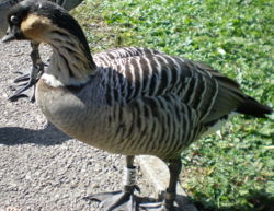
The beak, bill or rostrum of birds is formed from the bones of the maxilla and mandible with a horny, keratinised covering, the rhamphotheca. The beak is used for eating, grooming, manipulating objects, killing prey, probing for food, courtship and feeding their young. The morphology of the beak varies greatly from species to species and is mainly dependent on function.
Histologically, it is similar to the skin, with a modified epidermis. The stratum corneum is very thick, containing cell bound calcium phosphate and layered crystals of hydroxyapatite.
The beak is very sensitive to heat, cold, pressure and pain due to a high number of mechanoreceptors (Herbst corpuscles) being presence.
The corpuscles are recognisable histologically as papillae, originating from the dermis ending in crater-like structures at the distal tip of the beak.
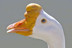
The cere is situated at the base of the upper beak and is composed of keratinised skin. The colour of the cere is influenced by diet and hormones.
The beak has two holes called nares (nostrils) which connect to the hollow inner beak and thence to the respiratory system. The nares are usually located on the dorsal beak. In some birds, they are located at the base of the beak in the cere.
In some species of bird, the tip of the beak is hard, dead tissue used for heavy-duty tasks such as cracking nuts or killing prey. In other species of bird, such as ducks, the tip of the bill is sensitive and contains nerves, for locating things by touch. The beak is worn down by use, so it grows continuously throughout the bird's life.
Reptiles
Reptilian Skin
The skin of reptiles has numerous functions including display, protection, camouflage, thermoregulation and fluid homeostasis.
The skin is dry, with few glands compared with mammals and amphibians. Glandular tissue is confined to femoral and precloacal pores in some lizards.
Epidermis
The epidermis consists of 3 layers:
- Stratum germinatum - which divides and produces keratin
- Stratum intermedium - which contains lipid, thus preventing fluid loss
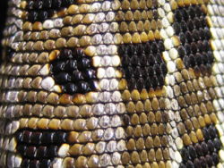
- Stratum corneum - which forms scales and scutes
In reptiles, 2 forms of keratin are present:
- Alpha-keratin which is flexible and often found between scales and scutes and in hinges
- Beta-keratin which is unique to reptiles. It is harder than alpha-keratin and forms scutes and scales
Dermis
The dermis of reptiles contains pigment cells, nerves and vessels, although thick, keratinised skin is without cutaneous sensation, leaving captive reptiles at risk of thermal burns. The dermis may contain bony plates called osteoderms for example, in the crocodile, tortoise and skink. The chelonian shell is formed from around 60 osteoderms which are fused with the ribs and parts of the spine and covered with epidermal scutes or leathery skin.
Pigment cells
- Chromatophores lie between the dermis and epidermis. They are influenced by the autonomic nervous system, hormones, light and temperature. They are used in camouflage, display and thermoregulation.
- Melanophores are related to the melanocytes of mammals and birds. They produce the colours black, brown, yellow and grey.
- Carotenoid cells are responsible for producing the colours red, yellow and orange.
- Iridophores (guanophores) lie in the dermis and contain the semi-crystalline product guanine that reflects light. Blue waves are reflected more, giving the skin a blue colour. When combined with yellow carotenoids, green colouring is formed.
Skin adaptations and cutaneous appendages
- Parietal eye - this is found in many lizards and is connected to the pineal gland. It is thought to be involved in thermoregulation.
- Spectacles - are clear scales over the eyes of snakes and geckos.
- Heat sensory pits (infrared-sensitive receptors) - are deep grooves between the nares and the eye of pit vipers, pythons and some boas which allow them to 'see' the radiated heat of their prey. These pits can also be found on the upper lip, just below the nares, termed 'labial pits'.
- Crests, frills, horns, gular pouches and spines - are used for display, and in the latter case, defence. There is often sexual dimorphism, with the appendages being larger in males.
- Cloacal spurs - are retained pelvic vestiges found in birds. They are generally used in courtship and are more pronounced in males than females.
- Rattle - present in some snakes and used to warn predators. It is enlarged with each shed.
- Gastropeges - are a single row of large ventral scales in snakes that aid locomotion.
- Adhesive toe-pads - are present in some geckos enabling them to grip surfaces including glass. They are composed of rows of tiny lamellae, each lamella, in turn, covered in tiny branching hairs called setae.
The Chelonian shell
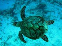
As previously mentioned, the shell comprises around 60 osteoderms which are fused with the ribs and part of the spine and covered by epidermal scutes or leathery skin.
The shell consists of a dome-shaped carapace dorsally and a flattened plastron ventrally. The exact size, shape and contour depends on the species. The pattern of the osteoderms and the overlying scutes do not match exactly, giving the shell additional strength. Scutes grow by the addition of new keratin layers to the base of each scute.
Ecdysis
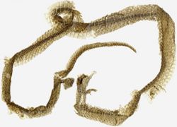
Ecdysis is the shedding of the skin in reptiles and is under the influence of the thyroid gland. Snakes and geckos tend to shed the whole skin and geckos also eat the shed skin, whereas other lizards shed piecemeal. Terrestrial tortoises shed legs, tail and neck skin only (piecemeal). Aquatic chelonia also shed individual scutes.
Stages of Ecdysis
- Stage 1: cells of the stratum intermedium replicate to form a new 3-layer epidermis.
- Stage 2: lymph and enzymes diffuse between the old and the new layers to form a cleavage zone.
- Stage 3: the old skin is then shed.
- Stage 4: the new skin hardens.
During ecdysis, the skin becomes more permeable and therefore more vulnerable to parasites and infection. Absorption of topical medications may also be enhanced, leading to toxicities. In snakes and some geckos, Stage 2 may most easily be recognised clinically as 'blue-white' discolouration of the spectacle. Snakes may become more aggressive and skittish at this time due to their temporarily impaired vision.
During ecdysis, many reptiles will seek out areas of increased humidity to facilitate proper shedding. Snakes especially may also require a rough object against which to rub to initiate final shedding. Failure to provide adequate humidity and/or a rough object, is a very common cause of dysecdysis in captive reptiles.
Amphibians
Amphibian skin
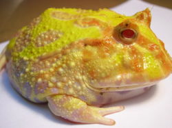
Amphibians are wholly or partly dependent on access to water and their integument is a very important organ. It functions as protection and as a sensory organ as well as having thermoregulatory and fluid homeostatic properties. Amphibian skin is very permeable, and many anurans (frogs and toads) have markedly increased vascularity over an area on the ventral pelvis known as the drinking patch or pelvic patch to enable water absorption (most amphibians don't drink).
Epidermis
The epidermis of amphibians is considerably thinner than that of reptiles and mammals and is easily damaged. The stratum corneum may be only 1 cell thick or may even be absent. Many amphibians shed and eat their skin on a regular basis.
Dermis
The dermis of amphibians consists of an outer stratum spongiosum and an inner stratum compactum. It contains nerves, vessels, smooth muscles, chromatophores and specialised glands.
In salamanders, the stratum compactum is tightly adhered to the underlying connective tissue.
In anurans (frogs and toads), the stratum compactum is loosly adhered to the underlying connective tissue providing a useful site for subcutaneous injection.
Glandular tissue
Glands in the epidermis may produce mucous or waxy substances which may serve to enhance cutaneous respiration and reduce evaporative water loss, respectively. Glands may also produce toxins and other chemicals which serve to protect against predators and infection.
Poison arrow frogs (Dendrobatids) are renowned for posessing potent toxins but only 2 or 3 species are known to produce enough toxin to be harmful to humans. Toxins are usually concentrated or metabolised from wild prey items and therefore captive bred and long-term captives produce little or no toxin, however, the importance of careful handling cannot be understressed.
Fire salamanders can spray toxin from dorsal glands.
Fish
Fish Skin
There are broadly 2 groups of fish: marine and freshwater fish. Marine fish have the tendency to gain salt and lose water, whereas freshwater fish tend to lose salt and gain water. The skin is a semi-waterproof barrier which helps maintain fluid and salt balance, in addition to the kidney and gills.
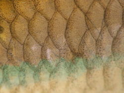
The skin consists of epidermal cells and scales (in most species) which are covered by a protective outer mucus cuticle. Most fish are covered in a protective layer of slime (mucus).
There are four types of fish scales.
- Placoid scales, also called dermal denticles, are similar to teeth in that they are made of dentin covered by enamel. They are typical of sharks and rays.
- Ganoid scales are flat, basal-looking scales that cover a fish body with little overlapping. They are typical of gar and bichirs.
- Cycloid scales are small oval-shaped scales with growth rings. Bowfin and remora have cycloid scales.
- Ctenoid scales are similar to the cycloid scales, with growth rings. They are distinguished by spines that cover one edge. Halibut have this type of scale.
Another, less common, type of scale is the scute, which is:
- An external shield-like bony plate
- A modified, thickened scale that often is keeled or spiny
- A projecting, modified (rough and strongly ridged) scale
- They are usually associated with the lateral line, or on the caudal peduncle forming caudal keels, or along the ventral profile.
Some fish, such as pineconefish, are completely or partially covered in scutes.
The epidermis and dermis of fish are similar to other vertebrates.
Epidermis
The epidermis consists of a stratum basale and a stratum germinatum. There is no stratum corneum or stratum spinosum though in some species (e.g. goldfish), accumulations of localised cornified cells occur during the breeding season ('breeding tubercles').
The thickness of the epidermis varies and contains mucus-secreting goblet cells. Some species have club cells which secrete alarm substances.
The cuticle consists of mucus and contains antibodies and lysosymes.
Unlike mammalian epidermal cells, fish epidermal cells at all levels are capable of cell division. During wound healing, cells migrate rapidly to cover any defect and restore the waterproof integrity of the skin.
Dermis
The dermis of fish consists of a stratum spongiosum and a deeper stratum compactum. It also contains chromatophores, mechanoreceptors, chemoreceptors and electroreceptors.
Scales are embedded in the dermis and covered by a layer of the epidermis; thus, the loss of scales will almost always damage the skin leading to osmotic balance problems.
Cutaneous appendages
- Specialised derivatives of scales include spines, stings, bony plates and the lateral line system.
- In fish, the lateral line is a sense organ used to detect movement and vibration in the surrounding water. Lateral lines are usually visible as faint lines running lengthways down each side, from the vicinity of the gill covers to the base of the tail. Sometimes parts of the lateral organ are modified into electroreceptors, which are organs used to detect electrical impulses. It is possible that vertebrates such as sharks use the lateral organs to detect magnetic fields as well. Most amphibian larvae and some adult amphibians also have a lateral organ.
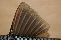
- Fins are modified structures of the skin that aid locomotion and balance in the water (and on land).
- Barbels are specialised sense organs around the mouth which contain many chemoreceptors
