Difference between revisions of "Gall Bladder - Anatomy & Physiology"
Jump to navigation
Jump to search
Fiorecastro (talk | contribs) |
|||
| (21 intermediate revisions by 5 users not shown) | |||
| Line 1: | Line 1: | ||
| − | {{ | + | {{toplink |
| + | |backcolour =BCED91 | ||
| + | |linkpage =Alimentary - Anatomy & Physiology | ||
| + | |linktext =Alimentary System | ||
| + | |maplink = | ||
| + | |pagetype =Anatomy | ||
| + | |sublink1=Liver - Anatomy & Physiology | ||
| + | |subtext1=THE LIVER | ||
| + | }} | ||
| + | <br> | ||
==Introduction== | ==Introduction== | ||
The gall bladder stores bile produced in the [[Liver - Anatomy & Physiology|liver]]. Bile is important in the digestion of lipids. | The gall bladder stores bile produced in the [[Liver - Anatomy & Physiology|liver]]. Bile is important in the digestion of lipids. | ||
| − | The gall bladder forms as an outgrowth of the bile duct, as a secondary hollow at the posterior edge of the original hepatic rudiment. The | + | The gall bladder forms as an outgrowth of the bile duct, as a secondary hollow at the posterior edge of the original hepatic rudiment. The gall bladder and the cyctic duct joins the common bile duct which enters the [[Duodenum - Anatomy & Physiology|duodenum]] at the major duodenal papillae (with the pancreatic duct) on the dorsal surface of the [[Duodenum - Anatomy & Physiology|duodenum]] |
==Structure== | ==Structure== | ||
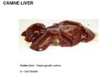
| − | [[Image:Canine Gallbladder Anatomy.jpg|thumb|right| | + | [[Image:Canine Gallbladder Anatomy.jpg|thumb|right|150px|Location of the Canine Gallbladder - Copyright RVC 2008]] |
| − | + | *Lies between the right medial and quadrate lobes of the [[Liver - Anatomy & Physiology|liver]] | |
| + | |||
| + | *Partly attached | ||
| + | |||
| + | *Partly free | ||
==Function== | ==Function== | ||
| − | + | *Stores bile | |
| + | |||
| + | *Concentrates bile by absorption through the folded mucosal wall | ||
==Innervation== | ==Innervation== | ||
| − | + | *Parasympathetic nerves | |
| + | |||
| + | ==Histology== | ||
| + | |||
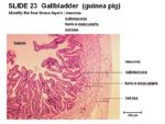
| + | [[Image:Guinea-pig Gallbladder Hsitology.jpg|thumb|right|150px|Histology of the Guinea-pig Gallbladder - Copyright RVC 2008]] | ||
| + | *Highly folded mucosa | ||
| + | |||
| + | *Reduced submucosa | ||
| + | |||
| + | *No lamina muscularis | ||
| + | |||
| + | *Simple columnar epithelium | ||
| + | |||
| + | *No glands | ||
==Species Differences== | ==Species Differences== | ||
'''Equine''' | '''Equine''' | ||
| − | + | *No gallbladder | |
'''Rodents''' | '''Rodents''' | ||
| − | + | *No gallbladder in rats | |
| − | |||
'''Canine''' | '''Canine''' | ||
| + | *Lies opposite the 8th intercostal space | ||
| − | + | *Thinest layers of tunica muscularis | |
'''Bovine''' | '''Bovine''' | ||
| + | *Thickest layers of tunica muscularis | ||
| + | |||
| + | *Sheep have a less projecting gall bladder than cows | ||
| − | + | *Gallbladder lies against the 10th or 11th rib | |
'''[[Avian Digestive Tract - Anatomy & Physiology|Avian]]''' | '''[[Avian Digestive Tract - Anatomy & Physiology|Avian]]''' | ||
| − | + | *Pigeons and parrots lack a gallbladder | |
| − | == | + | ==Test yourself with the Liver and Gallbladder flashcards== |
| − | [[ | + | [[Liver - Anatomy & Physiology - Flashcards|Liver & Gall Bladder Flashcards]] |
| − | |||
==Links== | ==Links== | ||
| − | + | [[Liver - gall bladder|Pathology of the Gall Bladder]] | |
| − | |||
| − | |||
| − | |||
| − | |||
| − | | | ||
| − | |||
| − | |||
| − | |||
| − | |||
| − | |||
| − | |||
| − | |||
Revision as of 14:45, 5 July 2010
|
|
Introduction
The gall bladder stores bile produced in the liver. Bile is important in the digestion of lipids.
The gall bladder forms as an outgrowth of the bile duct, as a secondary hollow at the posterior edge of the original hepatic rudiment. The gall bladder and the cyctic duct joins the common bile duct which enters the duodenum at the major duodenal papillae (with the pancreatic duct) on the dorsal surface of the duodenum
Structure
- Lies between the right medial and quadrate lobes of the liver
- Partly attached
- Partly free
Function
- Stores bile
- Concentrates bile by absorption through the folded mucosal wall
Innervation
- Parasympathetic nerves
Histology
- Highly folded mucosa
- Reduced submucosa
- No lamina muscularis
- Simple columnar epithelium
- No glands
Species Differences
Equine
- No gallbladder
Rodents
- No gallbladder in rats
Canine
- Lies opposite the 8th intercostal space
- Thinest layers of tunica muscularis
Bovine
- Thickest layers of tunica muscularis
- Sheep have a less projecting gall bladder than cows
- Gallbladder lies against the 10th or 11th rib
- Pigeons and parrots lack a gallbladder
Test yourself with the Liver and Gallbladder flashcards
Liver & Gall Bladder Flashcards

