Difference between revisions of "Snake Respiratory Disease"
| (8 intermediate revisions by 2 users not shown) | |||
| Line 9: | Line 9: | ||
History usually reveals inappropriate husbandry practices. Snakes with pneumonia will most often be presented when the disease is quite advanced. On [[Snake Physical Examination|physical examination]] dyspnoea may be seen. The respiration rate is generally elevated with exaggerated respiratory movements. Open-mouthed breathing is associated with serious pulmonary disease. A glottal discharge may be present as exudate moves from the lungs. The presence of nasal discharge is common with upper respiratory disease. On auscultation râles may be heard. | History usually reveals inappropriate husbandry practices. Snakes with pneumonia will most often be presented when the disease is quite advanced. On [[Snake Physical Examination|physical examination]] dyspnoea may be seen. The respiration rate is generally elevated with exaggerated respiratory movements. Open-mouthed breathing is associated with serious pulmonary disease. A glottal discharge may be present as exudate moves from the lungs. The presence of nasal discharge is common with upper respiratory disease. On auscultation râles may be heard. | ||
| + | '''For information on good husbandry practices, see''' [[:Category: Snake Husbandry|Snake Husbandry]]. | ||
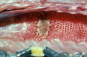
[[Image:Boiga_dendritica_Long_hhard.jpg|300px|thumb|right|'''The lung of a snake with pneumonia - A mycoplasma species has been isolated by PCR from respiratory tissue.''' © RVC]] | [[Image:Boiga_dendritica_Long_hhard.jpg|300px|thumb|right|'''The lung of a snake with pneumonia - A mycoplasma species has been isolated by PCR from respiratory tissue.''' © RVC]] | ||
| − | |||
==Diagnosis== | ==Diagnosis== | ||
Diagnosis of pneumonia is based on [[Lizard and Snake Taking a History|history]], [[Snake Physical Examination|physical examination]] and [[:Category: Snake Diagnostics|diagnostic aids]]. [[Lizard and Snake Imaging|Radiography]] is necessary for a definitive diagnosis of pneumonia. Lateral and dorsoventral views should be included. The pulmonary tissue normally has a delicate reticular pattern that is progressively lost caudally. Changes may be diffuse or regionalised. Samples for microbiology may be collected by glottal swabs, tracheal washes or endoscope. Cytology, gram stain and bacterial/fungal culture may help define the aetiology. Biopsy of lung tissue for histology may be diagnostic. | Diagnosis of pneumonia is based on [[Lizard and Snake Taking a History|history]], [[Snake Physical Examination|physical examination]] and [[:Category: Snake Diagnostics|diagnostic aids]]. [[Lizard and Snake Imaging|Radiography]] is necessary for a definitive diagnosis of pneumonia. Lateral and dorsoventral views should be included. The pulmonary tissue normally has a delicate reticular pattern that is progressively lost caudally. Changes may be diffuse or regionalised. Samples for microbiology may be collected by glottal swabs, tracheal washes or endoscope. Cytology, gram stain and bacterial/fungal culture may help define the aetiology. Biopsy of lung tissue for histology may be diagnostic. | ||
| − | '''For | + | '''For more information, see''' [[Snake Physical Examination]], [[Lizard and Snake Imaging]] '''and''' [[Lizard and Snake Specimen Collection and Evaluation]]. |
| − | |||
| − | |||
| − | |||
==Therapy== | ==Therapy== | ||
| − | A large percentage of respiratory disease has a bacterial component. Treatment should be based on culture and sensitivity but since respiratory disease is usually well advanced by the time of presentation, antibiotics should be commenced immediately. Enrofloxacin is a suitable antibiotic. Nebulisation may be helpful. Non-bacterial infections occur and therapy if possible should be aetiology-specific. Supportive treatment is extremely important. Keep all snakes within the upper end of their [[ | + | A large percentage of respiratory disease has a bacterial component. Treatment should be based on culture and sensitivity but since respiratory disease is usually well advanced by the time of presentation, antibiotics should be commenced immediately. Enrofloxacin is a suitable antibiotic. Nebulisation may be helpful. Non-bacterial infections occur and therapy if possible should be aetiology-specific. Supportive treatment is extremely important. Keep all snakes within the upper end of their [[POTZ]]. Fluid therapy and nutritional support are important but force-feeding should not stress the snake. Water-soluble vitamins may help. |
'''For more information, see''' [[Snake Supportive Care]]. | '''For more information, see''' [[Snake Supportive Care]]. | ||
| Line 28: | Line 25: | ||
'''For more information, see''' [[Lizard and Snake Quarantine]]. | '''For more information, see''' [[Lizard and Snake Quarantine]]. | ||
| − | |||
| − | |||
| − | |||
| − | |||
| − | |||
| − | |||
| − | |||
| − | |||
| − | |||
| − | |||
| − | |||
| − | |||
| − | |||
| − | |||
| − | |||
| − | |||
[[Category:Snake Diseases]] | [[Category:Snake Diseases]] | ||
Revision as of 08:55, 20 August 2010
| This article has been peer reviewed but is awaiting expert review. If you would like to help with this, please see more information about expert reviewing. |
Respiratory disease
Respiratory disease is common in snakes and respiratory signs are common presenting complaints. Pneumonia has a multifactorial aetiology including poor husbandry and infectious agents, usually Gram-negative bacteria.
For more information, see respiratory anatomy or physiology.
Examination
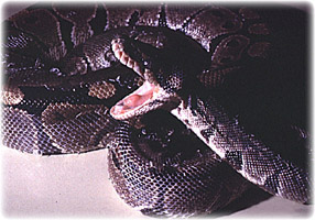
History usually reveals inappropriate husbandry practices. Snakes with pneumonia will most often be presented when the disease is quite advanced. On physical examination dyspnoea may be seen. The respiration rate is generally elevated with exaggerated respiratory movements. Open-mouthed breathing is associated with serious pulmonary disease. A glottal discharge may be present as exudate moves from the lungs. The presence of nasal discharge is common with upper respiratory disease. On auscultation râles may be heard.
For information on good husbandry practices, see Snake Husbandry.
Diagnosis
Diagnosis of pneumonia is based on history, physical examination and diagnostic aids. Radiography is necessary for a definitive diagnosis of pneumonia. Lateral and dorsoventral views should be included. The pulmonary tissue normally has a delicate reticular pattern that is progressively lost caudally. Changes may be diffuse or regionalised. Samples for microbiology may be collected by glottal swabs, tracheal washes or endoscope. Cytology, gram stain and bacterial/fungal culture may help define the aetiology. Biopsy of lung tissue for histology may be diagnostic.
For more information, see Snake Physical Examination, Lizard and Snake Imaging and Lizard and Snake Specimen Collection and Evaluation.
Therapy
A large percentage of respiratory disease has a bacterial component. Treatment should be based on culture and sensitivity but since respiratory disease is usually well advanced by the time of presentation, antibiotics should be commenced immediately. Enrofloxacin is a suitable antibiotic. Nebulisation may be helpful. Non-bacterial infections occur and therapy if possible should be aetiology-specific. Supportive treatment is extremely important. Keep all snakes within the upper end of their POTZ. Fluid therapy and nutritional support are important but force-feeding should not stress the snake. Water-soluble vitamins may help.
For more information, see Snake Supportive Care.
Prevention
Poor husbandry practices and non-hygienic conditions predispose to respiratory infections. Bacterial infections are often encountered in immunocompromised snakes. New arrivals into a collection should go through a period of quarantine.
For more information, see Lizard and Snake Quarantine.

