Difference between revisions of "Parotid Gland - Anatomy & Physiology"
Jump to navigation
Jump to search
Fiorecastro (talk | contribs) |
m (Text replace - "Category:Alimentary System" to "Category:Alimentary System - Anatomy & Physiology") |
||
| (19 intermediate revisions by 4 users not shown) | |||
| Line 1: | Line 1: | ||
| + | {{toplink | ||
| + | |linkpage =Alimentary - Anatomy & Physiology | ||
| + | |linktext =Alimentary System | ||
| + | |maplink = | ||
| + | |sublink1=Oral Cavity - Salivary Glands - Anatomy & Physiology#Major Salivary Glands | ||
| + | |subtext1=MAJOR SALIVARY GLANDS | ||
| + | }} | ||
| + | <br> | ||
| + | ==Parotid Salivary Gland== | ||
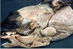
| + | [[Image:Parotid & Mandibular Salivary Gland.jpg|thumb|right|150px|Parotid Salivary Gland - Copyright Nottingham 2008]] | ||
| + | *[[Serous Salivary Gland - Anatomy and Physiology|Serous]] secretion | ||
| − | + | *Moulded around base of [[Ear - Anatomy & Physiology#Outer Ear|auricular cartilage]] of ear | |
| − | [[ | ||
| − | + | *Enclosed within facial covering | |
| − | Trabeculae divide | + | *Trabeculae divide gland into lobules |
| − | + | *Major ducts run within trabeculae and merge to form a single duct | |
| + | |||
| + | *Duct opens in vestibule opposite 4th upper premolar (Not all species) | ||
| + | |||
| + | *Innervated by glossopharyngeal ([[Cranial Nerves - Anatomy & Physiology|CN IX]]) via trigeminal branch ([[Cranial Nerves - Anatomy & Physiology|CN V]]) | ||
==Development== | ==Development== | ||
| − | Intercalated duct | + | 1. Intercalated duct. Cuboidal cells. |
| + | |||
| + | 2. Striated duct. Cuboidal cells with mitochondria in base. | ||
| + | |||
| + | 3. Interlobular duct. Columnar to stratified columnar cells. | ||
| + | |||
| + | 4. Stratified squamous epithelium continuous with epithelium lining [[Oral Cavity Overview - Anatomy & Physiology|oral cavity]] | ||
| + | |||
==Histology== | ==Histology== | ||
| − | + | *Basophilic endoplasmic reticulum | |
| + | |||
| + | *Stratified squamous epithelium | ||
| + | |||
| + | *Tubulo-acinar gland | ||
| + | |||
| + | *Acinar cells surrounded by myoepithelial cells and basement membrane | ||
| + | |||
==Species Differences== | ==Species Differences== | ||
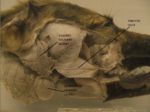
| + | [[Image:Parotid Duct.jpg|thumb|right|150px|Parotid Duct (Dog) - Copyright RVC]] | ||
| + | ===Carnivores=== | ||
| + | *Some mucous secretion in cat and dog | ||
| − | + | *Duct is superficial in dog | |
| − | + | *Duct runs across masseter muscle in carnivores | |
===Herbivores=== | ===Herbivores=== | ||
| + | *Larger gland and higher flow rate in herbivores to lubricate and soften food | ||
| − | + | *Duct is superficial in small ruminants | |
| + | |||
| + | *Parotid gland extends rostrally over masseter muscle, ventrally to angle of jaw and caudally towards atlantal fossa | ||
| + | |||
| + | *Duct runs ventrally in herbivores below the [[Skull and Facial Muscles - Anatomy & Physiology#Mandibule (mandibula)|mandible]] (facial groove in horses) before entering the [[Oral Cavity Overview - Anatomy & Physiology|oral cavity]] at the rostral margin of the masseter muscle | ||
===Equine=== | ===Equine=== | ||
| − | + | *Gland overlies the [[Ear - Anatomy & Physiology#Equine Gutteral Pouch|gutteral pouch]] | |
| − | |||
<br /> | <br /> | ||
<br /> | <br /> | ||
| + | ==Test yourself with the salivary glands flashcards== | ||
| + | |||
| + | [[Oral_Cavity_-_Anatomy_%26_Physiology_-_Flashcards#Salivary_Glands_Flashcards|Salivary Glands Flashcards]] | ||
| + | |||
| + | ==Links== | ||
| − | + | '''Video''' | |
| − | + | [http://stream2.rvc.ac.uk/Anatomy/canine/head_neck/Pot0258.mp4 Pot 258 Lateral section through the head of a dog] | |
| − | |||
| − | |||
| − | |||
| − | |||
| − | |||
| − | [[Category: | + | [[Category:Alimentary System - Anatomy & Physiology]] |
| − | |||
Revision as of 15:32, 31 August 2010
|
|
Parotid Salivary Gland
- Serous secretion
- Moulded around base of auricular cartilage of ear
- Enclosed within facial covering
- Trabeculae divide gland into lobules
- Major ducts run within trabeculae and merge to form a single duct
- Duct opens in vestibule opposite 4th upper premolar (Not all species)
Development
1. Intercalated duct. Cuboidal cells.
2. Striated duct. Cuboidal cells with mitochondria in base.
3. Interlobular duct. Columnar to stratified columnar cells.
4. Stratified squamous epithelium continuous with epithelium lining oral cavity
Histology
- Basophilic endoplasmic reticulum
- Stratified squamous epithelium
- Tubulo-acinar gland
- Acinar cells surrounded by myoepithelial cells and basement membrane
Species Differences
Carnivores
- Some mucous secretion in cat and dog
- Duct is superficial in dog
- Duct runs across masseter muscle in carnivores
Herbivores
- Larger gland and higher flow rate in herbivores to lubricate and soften food
- Duct is superficial in small ruminants
- Parotid gland extends rostrally over masseter muscle, ventrally to angle of jaw and caudally towards atlantal fossa
- Duct runs ventrally in herbivores below the mandible (facial groove in horses) before entering the oral cavity at the rostral margin of the masseter muscle
Equine
- Gland overlies the gutteral pouch

