Difference between revisions of "African Horse Sickness"
| (3 intermediate revisions by the same user not shown) | |||
| Line 1: | Line 1: | ||
| + | {{OpenPagesTop}} | ||
== Introduction == | == Introduction == | ||
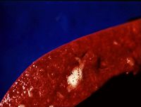
| + | [[Image:Lung oedema in African horse sickness.jpg|thumb|right|200px|<small><center>Lung oedema in African horse sickness (Image sourced from Bristol Biomed Image Archive with permission)</center></small>]] | ||
| + | This is a serious viral disease transmitted by [[Ceratopogonidae|midges]] causing systemic signs and often death in the horse. It is a worldwide disease but is currently not found in the UK and is notifiable here. However, with global warming, the midge breeding area is growing and there are worries that in the future the disease will occur here. It affects horses, donkeys and mules and also is present subclinically in zebras and african donkeys. These can be a reservoir of infection for the disease. The disease has only been found in Africa, but since 1987 has recurred in Spain and Portugal. | ||
| − | + | There are nine serotypes of African Horse Sickness. The vectors for the disease are [[Ceratopogonidae|'''midges''']]. The virus oscillates between horses and midges. It replicates in the salivary glands of biting midges, and occurs where they breed. | |
| − | + | The virus is pantropic and infects the endothelium and myocardium. It enters via the bloodstream from a midge bite and then disseminates throughout the body. | |
| − | |||
| − | + | == Clinical Signs== | |
| − | |||
| − | == Clinical Signs | ||
'''Peracute''': causes sudden death with pyrexia before the onset of clinical signs. | '''Peracute''': causes sudden death with pyrexia before the onset of clinical signs. | ||
| − | '''Acute | + | '''Acute cardiac-pneumonic form''':causes signs such as interlobular [[Pulmonary Oedema|pulmonary oedema]], [[pericarditis]], hemorrhage and [[Oedema|oedema]] of the viscera. Death usually occurs within 5 days. |
| − | '''Subacute cardiac form''': causes milder clinical signs but still a high mortality rate. | + | '''Subacute cardiac form''': causes milder clinical signs but still a high mortality rate. |
| − | |||
| − | == Diagnosis | + | == Diagnosis== |
| − | Clinical signs are quite presumptive and would lead you to be concerned of a severe viral infection and inflict necessary precautions such as isolation etc. Definitive diagnosis should be made via | + | Clinical signs are quite presumptive and would lead you to be concerned of a severe viral infection and inflict necessary precautions such as isolation etc. Definitive diagnosis should be made via elimination of differential diagnoses such as [[Equine Arteritis Virus]] (EAV). EAV causes oedema in the legs, which AHS does NOT. |
Samples should be taken for virus serotype identification and serology. | Samples should be taken for virus serotype identification and serology. | ||
| − | |||
| − | == Treatment and Control | + | == Treatment and Control == |
| − | In countries where the disease is not endemic, e.g. the UK, then humane destruction of the horse will be required. | + | In countries where the disease is not endemic, e.g. the UK, then humane destruction of the horse will be required. |
Control measures are largely preventative and include slaughter or immediate isolation of sick animals, mandatory and immediate vaccination with appropriate serotype, restricted movement of susceptible animals and protection zones and surveillance zones set in place around outbreaks. The control of the vector with ectoparasiticides, etc is also very important. | Control measures are largely preventative and include slaughter or immediate isolation of sick animals, mandatory and immediate vaccination with appropriate serotype, restricted movement of susceptible animals and protection zones and surveillance zones set in place around outbreaks. The control of the vector with ectoparasiticides, etc is also very important. | ||
| − | |||
| − | == References | + | == References == |
Reed, S.M, Bayly, W.M. and Sellon, D.C (2010) Equine Internal Medicine (Third Edition), Saunders. <br>Reed, S.M, Bayly, W.M, Sellon, D.C. (2004) Equine Internal Medicine (Second Edition) Saunders. <br>Robinson, N.E., Sprayberry, K.A. (2009) Current Therapy in Equine Medicine (Sixth Edition) Saunders Elsevier<br>Rose, R. J. and Hodgson, D. R. (2000) Manual of Equine Practice (Second Edition) Sauders. <br> | Reed, S.M, Bayly, W.M. and Sellon, D.C (2010) Equine Internal Medicine (Third Edition), Saunders. <br>Reed, S.M, Bayly, W.M, Sellon, D.C. (2004) Equine Internal Medicine (Second Edition) Saunders. <br>Robinson, N.E., Sprayberry, K.A. (2009) Current Therapy in Equine Medicine (Sixth Edition) Saunders Elsevier<br>Rose, R. J. and Hodgson, D. R. (2000) Manual of Equine Practice (Second Edition) Sauders. <br> | ||
| − | |||
| − | + | {{review}} | |
| − | + | {{OpenPages}} | |
| − | [[Category:Orbiviruses]] [[Category:Horse_Viruses]] [[Category: | + | [[Category:Orbiviruses]] [[Category:Horse_Viruses]] [[Category:Expert_Review - Horse]] [[Category:Respiratory_Viral_Infections]] [[Category:Respiratory_Diseases_-_Horse]] |
Latest revision as of 16:54, 31 July 2012
Introduction
This is a serious viral disease transmitted by midges causing systemic signs and often death in the horse. It is a worldwide disease but is currently not found in the UK and is notifiable here. However, with global warming, the midge breeding area is growing and there are worries that in the future the disease will occur here. It affects horses, donkeys and mules and also is present subclinically in zebras and african donkeys. These can be a reservoir of infection for the disease. The disease has only been found in Africa, but since 1987 has recurred in Spain and Portugal.
There are nine serotypes of African Horse Sickness. The vectors for the disease are midges. The virus oscillates between horses and midges. It replicates in the salivary glands of biting midges, and occurs where they breed.
The virus is pantropic and infects the endothelium and myocardium. It enters via the bloodstream from a midge bite and then disseminates throughout the body.
Clinical Signs
Peracute: causes sudden death with pyrexia before the onset of clinical signs.
Acute cardiac-pneumonic form:causes signs such as interlobular pulmonary oedema, pericarditis, hemorrhage and oedema of the viscera. Death usually occurs within 5 days.
Subacute cardiac form: causes milder clinical signs but still a high mortality rate.
Diagnosis
Clinical signs are quite presumptive and would lead you to be concerned of a severe viral infection and inflict necessary precautions such as isolation etc. Definitive diagnosis should be made via elimination of differential diagnoses such as Equine Arteritis Virus (EAV). EAV causes oedema in the legs, which AHS does NOT.
Samples should be taken for virus serotype identification and serology.
Treatment and Control
In countries where the disease is not endemic, e.g. the UK, then humane destruction of the horse will be required.
Control measures are largely preventative and include slaughter or immediate isolation of sick animals, mandatory and immediate vaccination with appropriate serotype, restricted movement of susceptible animals and protection zones and surveillance zones set in place around outbreaks. The control of the vector with ectoparasiticides, etc is also very important.
References
Reed, S.M, Bayly, W.M. and Sellon, D.C (2010) Equine Internal Medicine (Third Edition), Saunders.
Reed, S.M, Bayly, W.M, Sellon, D.C. (2004) Equine Internal Medicine (Second Edition) Saunders.
Robinson, N.E., Sprayberry, K.A. (2009) Current Therapy in Equine Medicine (Sixth Edition) Saunders Elsevier
Rose, R. J. and Hodgson, D. R. (2000) Manual of Equine Practice (Second Edition) Sauders.
| This article has been peer reviewed but is awaiting expert review. If you would like to help with this, please see more information about expert reviewing. |
Error in widget FBRecommend: unable to write file /var/www/wikivet.net/extensions/Widgets/compiled_templates/wrt695ce49abae2e2_00611572 Error in widget google+: unable to write file /var/www/wikivet.net/extensions/Widgets/compiled_templates/wrt695ce49ac0b584_78012009 Error in widget TwitterTweet: unable to write file /var/www/wikivet.net/extensions/Widgets/compiled_templates/wrt695ce49ac71c83_35508282
|
| WikiVet® Introduction - Help WikiVet - Report a Problem |
