Difference between revisions of "Oesophageal Stricture"
TestStudent (talk | contribs) |
|||
| (27 intermediate revisions by 4 users not shown) | |||
| Line 1: | Line 1: | ||
| − | {{ | + | {{OpenPagesTop}} |
| − | + | ==Introduction== | |
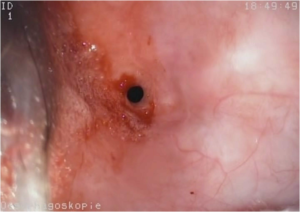
| − | + | [[Image:Oesophageal Stricture.png|thumb|right|300px|An oesophageal stricture observed by oesophagoscopy<br><small> Copyright David Walker 2009 RVC]]</small> | |
| − | + | An oesophageal stricture is an abnormal circumferential narrowing of the oesophageal lumen secondary to severe [[Oesophagitis|oesophagitis]]. Deep injuries to the oesophageal wall heal by fibrosis which contracts over time to form the stricture. Animals may suffer from multiple strictures and the most common sites are within the '''distal high pressure zone''' (close to the lower oesophageal spincter), over the '''base of the heart''' and '''at the thoracic inlet'''. The oesophagus is narrower at these points and foreign bodies and refluxed ingesta are therefore more likely to accumulate at these locations. The most important causes of strictures include: | |
| − | + | *'''Physical Injury''' | |
| − | == | + | **Ingestion of [[Oesophageal Foreign Body|'''foreign bodies''']] which lodge in the oesophagus. |
| − | * | + | **Passage of nasogastric or pharyngostomy '''feeding tubes''' or of large '''endoscopes'''. |
| − | * | + | **Erosion of the oesophageal wall or extraluminal compression by '''neoplasia''' or '''abscesses'''. |
| − | * | + | **'''Surgical wounds''' to the oesophagus that heal with a fibrous component. |
| − | * | + | *'''Chemical Injury''' |
| + | **'''Gastro-oesophageal reflux''', which may occur with '''general anaesthesia''' or [[Hiatal Hernia|'''hiatal hernias''']]. | ||
| + | **'''Chronic vomiting''' | ||
| + | **Ingestion of '''caustic''' or '''irritant substances''', including '''doxycycline''' in cats. | ||
| − | == | + | ==Signalment== |
| − | + | The signalment is dependent on the underlying cause of the injury, with animals of all ages having the potential to be affected. 46% of cases occur after general anaesthesia, presumably due to gastro-oesophageal reflux through the relaxed lower oesophageal sphincter and because peristalsis and saliva production are reduced, meaning that refluxed material is not removed or neutralised. | |
| − | |||
| − | |||
| − | |||
| − | |||
| − | |||
| − | |||
==Diagnosis== | ==Diagnosis== | ||
| − | |||
===Clinical Signs=== | ===Clinical Signs=== | ||
| − | + | The signs observed depend on the severity and extent of the stricture but include: | |
| − | *Regurgitation shortly after feeding | + | *'''Regurgitation''' shortly after feeding, often with '''hypersalivation/ptyalism'''. Liquid foods may be better tolerated than solid food and it is better able to pass through the stricture. |
| − | *Anorexia | + | *'''Anorexia''' and resultant '''weight loss'''. |
| − | + | *As with any cause of repeated regurgitation, '''aspiration pneumonia''' may develop with signs of coughing, tachypnoea, dyspnoea and pyrexia. | |
| − | |||
| − | |||
| − | * | ||
| − | |||
| − | |||
| − | |||
===Diagnostic Imaging=== | ===Diagnostic Imaging=== | ||
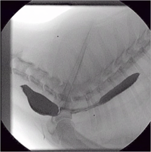
| − | Fibrosing strictures must be differentiated from vascular ring anomalies, [[Oesophagitis | + | [[Image:Oesophageal Stricture Flouroscopy.png|thumb|right|300px|An oesophageal stricture observed by fluoroscopy in the region of the thoracic inlet <br><small> Copyright David Walker 2009 RVC]]</small> |
| + | Fibrosing strictures must be differentiated from [[Vascular Ring Anomalies|vascular ring anomalies]], [[Oesophagitis|oesophagitis]] and intraluminal and extraluminal masses. This can be done with plain and contrast radiography, endoscopy and ultrasonography. | ||
| − | + | '''Plain radiographs''' are usually unremarkable in animals with simple oesophageal strictures but oral administration of '''barium contrast medium''' may demonstrate '''segmental''' or '''diffuse narrowing''' of the oesophagus and '''oesphageal dilatation''' proximal to the site of the stricture. | |
| − | |||
| − | |||
| − | + | '''Fluoroscopy''' can be used to great effect to observe the passage of a food bolus along the oesophagus and to define the sites of multiple strictures. | |
| − | Oesophagoscopy | + | '''Ultrasonography''' is not usually useful in diagnosing simple strictures but may visualise those caused by extraluminal compression by mediastinal neoplasia or abscesses. |
| + | |||
| + | '''Oesophagoscopy''' may be used to obtain a definitive diagnosis but it requires a general anaesthetic (which may have been the cause of the problem) and, if a large endoscope is passed, this may cause further physical damage to the oesophagus. Also, endoscopy is not sensitive for the diagnosis of multiple strictures if it is unable to pass through the most proximal. It can also be used to identify and biopsy any intraluminal masses. | ||
==Treatment== | ==Treatment== | ||
| + | ===Medical Management=== | ||
| + | The suspected cause (ie.[[Oesophagitis|oesophagitis]]) should be corrected initially. Components of medical management include: | ||
| + | *'''Withdrawal of oral food for 2-3 days''' as standard but, if the inflammation is severe or rupture has occurred, a '''gastrostomy tube''' may be required. | ||
| + | *'''Oral sucralfate suspension''' is thought to bind to the base of any ulcers, to stimulate epithelial repair and to neutralise any refluxed gastric juices. | ||
| + | *'''Gastric acid secretory inhibitors''' (e.g. ranitidine, omeprazole) can be useful in cases of gastro-oesophageal reflux. | ||
| + | *'''Metaclopramide''', a promotility drug that increases the tone of the lower oesophageal sphincter, may also be used to manage gastro-oesophageal reflux but not if oesophageal motility is thought to be impaired (i.e., if megaoesophagus is present). | ||
| + | *'''Broad spectrum intra-venous bactericidal antibiotics''' may be required in animals with severe oesophagitis or aspiration pneumonia. Coupage and nebulisation may be useful adjunctive treatments for aspiration pneumonia and it may be necessary to provide oxygen to dyspnoeic animals. | ||
| + | *'''Analgesia''' should be provided to encourage animals to eat after 2-3 days. | ||
| + | *Anti-inflammatory doses of [[Steroids|corticosteroids]] (such as prednisolone) may be used to prevent fibrosis and further stricture formation in acute injuries but caution should be exercised if the animal has concurrent aspiration pneumonia. | ||
| + | |||
| + | ===Surgical Management=== | ||
| + | Surgical intervention usually involves gradually stretching the site of the stricture until the luminal diameter returns to normal. Since flow increases as the fourth power of the radius (according to the law of LaPlace), even small increases in diameter can produce significant improvements in clinical signs. Multiple procedures (4-12) are usually required to achieve acceptable results. Bougeinage and balloon catheters are the methods used most commonly. | ||
| − | + | '''Bougeinage''' involves the passage of conical metal rods of increasing size along the oesophagus but alternatively, a '''balloon catheter''' can be advanced along and oesophagus and inflated at the site of the stricture. This technique exerts pressure more evenly on the entire stricture, especially if it is not circumferential. | |
| − | |||
| − | + | '''Surgical resection and anastomosis''' is not recommended because iatrogenic strictures may form at the anastomotic site. A simpler '''oesophagoplasty''' (similar to pyloroplasty for pyloric stenosis) may be performed, where the stricture is incised longitudinally and sutured transversely to increase the luminal diameter. | |
| − | |||
| − | |||
| − | |||
| − | + | Anti-inflammatory doses of corticosteroids should again be used during these procedures to try to prevent re-stricturing. | |
| − | |||
| − | |||
==Prognosis== | ==Prognosis== | ||
| − | The shorter the length of oesophagus involved and the quicker the corrective procedure is performed the better the prognosis. | + | The shorter the length of oesophagus involved and the quicker the corrective procedure is performed, the better the prognosis. Animals with large, mature strictures and those with continued oesophagitis have a guarded prognosis and long-term gastrostomy tubes may be required in these cases. |
| − | Animals with large, mature strictures and those with continued oesophagitis have a guarded prognosis | + | |
| + | {{Learning | ||
| + | |Vetstream = [https://www.vetstream.com/canis/Content/Disease/dis00639.asp, Oesophageal stricture] | ||
| + | |literature search = [http://www.cabdirect.org/search.html?q=%28title%3A%28oesophag*%29+OR+title%3A%28esophag*%29%29+AND+title%3A%28stricture%29 Oesophageal Stricture publications] | ||
| + | }} | ||
==References== | ==References== | ||
*Hall, E.J, Simpson, J.W. and Williams, D.A. (2005) '''BSAVA Manual of Canine and Feline Gastroenterology (2nd Edition)''' ''BSAVA'' | *Hall, E.J, Simpson, J.W. and Williams, D.A. (2005) '''BSAVA Manual of Canine and Feline Gastroenterology (2nd Edition)''' ''BSAVA'' | ||
| + | *Merck & Co (2008) '''The Merck Veterinary Manual'''''Merial | ||
| + | *Nelson, R.W. and Couto, C.G. (2009) '''Small Animal Internal Medicine (Fourth Edition)''' ''Mosby Elsevier''. | ||
| + | |||
| + | |||
| + | {{review}} | ||
| − | + | {{OpenPages}} | |
| − | + | [[Category:Oesophagus_-_Pathology]] | |
| + | [[Category:Expert_Review - Small Animal]] | ||
| + | [[Category:Oesophageal Diseases - Dog]][[Category:Oesophageal Diseases - Cat]] | ||
Latest revision as of 18:56, 1 September 2015
Introduction
An oesophageal stricture is an abnormal circumferential narrowing of the oesophageal lumen secondary to severe oesophagitis. Deep injuries to the oesophageal wall heal by fibrosis which contracts over time to form the stricture. Animals may suffer from multiple strictures and the most common sites are within the distal high pressure zone (close to the lower oesophageal spincter), over the base of the heart and at the thoracic inlet. The oesophagus is narrower at these points and foreign bodies and refluxed ingesta are therefore more likely to accumulate at these locations. The most important causes of strictures include:
- Physical Injury
- Ingestion of foreign bodies which lodge in the oesophagus.
- Passage of nasogastric or pharyngostomy feeding tubes or of large endoscopes.
- Erosion of the oesophageal wall or extraluminal compression by neoplasia or abscesses.
- Surgical wounds to the oesophagus that heal with a fibrous component.
- Chemical Injury
- Gastro-oesophageal reflux, which may occur with general anaesthesia or hiatal hernias.
- Chronic vomiting
- Ingestion of caustic or irritant substances, including doxycycline in cats.
Signalment
The signalment is dependent on the underlying cause of the injury, with animals of all ages having the potential to be affected. 46% of cases occur after general anaesthesia, presumably due to gastro-oesophageal reflux through the relaxed lower oesophageal sphincter and because peristalsis and saliva production are reduced, meaning that refluxed material is not removed or neutralised.
Diagnosis
Clinical Signs
The signs observed depend on the severity and extent of the stricture but include:
- Regurgitation shortly after feeding, often with hypersalivation/ptyalism. Liquid foods may be better tolerated than solid food and it is better able to pass through the stricture.
- Anorexia and resultant weight loss.
- As with any cause of repeated regurgitation, aspiration pneumonia may develop with signs of coughing, tachypnoea, dyspnoea and pyrexia.
Diagnostic Imaging
Fibrosing strictures must be differentiated from vascular ring anomalies, oesophagitis and intraluminal and extraluminal masses. This can be done with plain and contrast radiography, endoscopy and ultrasonography.
Plain radiographs are usually unremarkable in animals with simple oesophageal strictures but oral administration of barium contrast medium may demonstrate segmental or diffuse narrowing of the oesophagus and oesphageal dilatation proximal to the site of the stricture.
Fluoroscopy can be used to great effect to observe the passage of a food bolus along the oesophagus and to define the sites of multiple strictures.
Ultrasonography is not usually useful in diagnosing simple strictures but may visualise those caused by extraluminal compression by mediastinal neoplasia or abscesses.
Oesophagoscopy may be used to obtain a definitive diagnosis but it requires a general anaesthetic (which may have been the cause of the problem) and, if a large endoscope is passed, this may cause further physical damage to the oesophagus. Also, endoscopy is not sensitive for the diagnosis of multiple strictures if it is unable to pass through the most proximal. It can also be used to identify and biopsy any intraluminal masses.
Treatment
Medical Management
The suspected cause (ie.oesophagitis) should be corrected initially. Components of medical management include:
- Withdrawal of oral food for 2-3 days as standard but, if the inflammation is severe or rupture has occurred, a gastrostomy tube may be required.
- Oral sucralfate suspension is thought to bind to the base of any ulcers, to stimulate epithelial repair and to neutralise any refluxed gastric juices.
- Gastric acid secretory inhibitors (e.g. ranitidine, omeprazole) can be useful in cases of gastro-oesophageal reflux.
- Metaclopramide, a promotility drug that increases the tone of the lower oesophageal sphincter, may also be used to manage gastro-oesophageal reflux but not if oesophageal motility is thought to be impaired (i.e., if megaoesophagus is present).
- Broad spectrum intra-venous bactericidal antibiotics may be required in animals with severe oesophagitis or aspiration pneumonia. Coupage and nebulisation may be useful adjunctive treatments for aspiration pneumonia and it may be necessary to provide oxygen to dyspnoeic animals.
- Analgesia should be provided to encourage animals to eat after 2-3 days.
- Anti-inflammatory doses of corticosteroids (such as prednisolone) may be used to prevent fibrosis and further stricture formation in acute injuries but caution should be exercised if the animal has concurrent aspiration pneumonia.
Surgical Management
Surgical intervention usually involves gradually stretching the site of the stricture until the luminal diameter returns to normal. Since flow increases as the fourth power of the radius (according to the law of LaPlace), even small increases in diameter can produce significant improvements in clinical signs. Multiple procedures (4-12) are usually required to achieve acceptable results. Bougeinage and balloon catheters are the methods used most commonly.
Bougeinage involves the passage of conical metal rods of increasing size along the oesophagus but alternatively, a balloon catheter can be advanced along and oesophagus and inflated at the site of the stricture. This technique exerts pressure more evenly on the entire stricture, especially if it is not circumferential.
Surgical resection and anastomosis is not recommended because iatrogenic strictures may form at the anastomotic site. A simpler oesophagoplasty (similar to pyloroplasty for pyloric stenosis) may be performed, where the stricture is incised longitudinally and sutured transversely to increase the luminal diameter.
Anti-inflammatory doses of corticosteroids should again be used during these procedures to try to prevent re-stricturing.
Prognosis
The shorter the length of oesophagus involved and the quicker the corrective procedure is performed, the better the prognosis. Animals with large, mature strictures and those with continued oesophagitis have a guarded prognosis and long-term gastrostomy tubes may be required in these cases.
| Oesophageal Stricture Learning Resources | |
|---|---|
To reach the Vetstream content, please select |
Canis, Felis, Lapis or Equis |
 Search for recent publications via CAB Abstract (CABI log in required) |
Oesophageal Stricture publications |
References
- Hall, E.J, Simpson, J.W. and Williams, D.A. (2005) BSAVA Manual of Canine and Feline Gastroenterology (2nd Edition) BSAVA
- Merck & Co (2008) The Merck Veterinary ManualMerial
- Nelson, R.W. and Couto, C.G. (2009) Small Animal Internal Medicine (Fourth Edition) Mosby Elsevier.
| This article has been peer reviewed but is awaiting expert review. If you would like to help with this, please see more information about expert reviewing. |
Error in widget FBRecommend: unable to write file /var/www/wikivet.net/extensions/Widgets/compiled_templates/wrt6759240cc54145_28956680 Error in widget google+: unable to write file /var/www/wikivet.net/extensions/Widgets/compiled_templates/wrt6759240ccc8ac4_18785133 Error in widget TwitterTweet: unable to write file /var/www/wikivet.net/extensions/Widgets/compiled_templates/wrt6759240cd1c1b4_93369129
|
| WikiVet® Introduction - Help WikiVet - Report a Problem |

