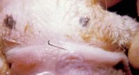
Bovine Papular Stomatitis
Introduction
This disease is caused by a Parapox virus and is a very similar disease to orf but seen in cattle and is generally a milder condition. It must be differentiated from Foot and Mouth Disease and Mucosal Disease.
It tends to occur sporadically in cattle that are less than one year of age. Lesions develop on the muzzle, external nares and in the oral cavity; the oesophagus and forestomachs may also be affected. A characteristic feature of the disease is that it usually heals spontaneously.
The early lesions are round areas of intense congestion up to 1.5 cm in diameter and then the centre becomes necrotic and slightly depressed. Slow peripheral extension of this lesion gives a classical ring zone formation with concentric rings of yellow (necrosis), grey (epithelial hyperplasia) and red (congestion).
Signalment
Young cattle; less than one year old
Clinical Signs
Lesions around the muzzle, nares and in the oral cavity that have a typical 'pock-like' appearance. The animal will be otherwise completely well. If the lesions are in the oral cavity, it may at first have difficulty eating, but not so much so that there will be any noticeable signs of weight loss.
Diagnosis
Clinical signs are characteristic, but the disease needs to be differentiated from Foot and Mouth and Mucosal disease, which with thorough inspection of lesions should be easy as vesicle formation is not a feature of Bovine Papular Stomatitis.
Diagnosis is usually made by clinical signs, but if a biopsy were to be taken of the lesion one would see focal areas of hydropic degeneration in the stratum spinosum, large eosinophilic intracytoplasmic inclusions, the epidermis will appear markedly thickened and the superficial layers of the epithelium become necrotic and slough.
Control
There are no vaccines available and treatment is not necessary as the lesions resolve spontaneously.
| Bovine Papular Stomatitis Learning Resources | |
|---|---|
To reach the Vetstream content, please select |
Canis, Felis, Lapis or Equis |
References
Andrews, A.H, Blowey, R.W, Boyd, H and Eddy, R.G. (2004) Bovine Medicine (Second edition), Blackwell Publishing
Radostits, O.M, Arundel, J.H, and Gay, C.C. (2000) Veterinary Medicine: a textbook of the diseases of cattle, sheep, pigs, goats and horses, Elsevier Health Sciences
| This article has been peer reviewed but is awaiting expert review. If you would like to help with this, please see more information about expert reviewing. |
Error in widget FBRecommend: unable to write file /var/www/wikivet.net/extensions/Widgets/compiled_templates/wrt695a514db08d05_22962241 Error in widget google+: unable to write file /var/www/wikivet.net/extensions/Widgets/compiled_templates/wrt695a514db75130_75262057 Error in widget TwitterTweet: unable to write file /var/www/wikivet.net/extensions/Widgets/compiled_templates/wrt695a514dbe4991_05297432
|
| WikiVet® Introduction - Help WikiVet - Report a Problem |

