Ventricular Septal Defect
Introduction
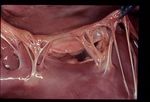
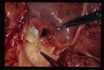
Ventricular septal defects (VSDs) are the most common congenital cardiac abnormality in large animals and the second most common congenital cardiac abnormality in cats (12-56% of congenital heart disease cases). They less commonly occur in dogs (6-12% of congenital heart disease cases).
The lesion is usually in the membranous area of the interventricular septum, just below the aortic valve and the septal tricuspid leaflet.
Ventricular Septal Defects can occur alone or in combination with other congenital malformations, such as Tetralogy of Fallot. Usually blood flows from the higher pressure left ventricle through the VSD to the lower pressure right ventricle during systole. Significant left-to-right shunting causes volume overload of the pulmonary circulation, increasing pulmonary venous return to the left atrium and consequently the left ventricle. This may result in volume overload of the left heart with consequent left-sided congestive heart failure and pulmonary hypertension. If chronic over-circulation of the pulmonary vasculature occurs, pulmonary hypertension may develop. Pulmonary hypertension causes increased right ventricular pressures and therefore right-to-left shunting during systole may occur; this is known as Eisenmenger's syndrome. Right-to-left shunting allows deoxygenated blood to enter the systemic circulation, resulting in arterial hypoxaemia and cyanosis.
Small VSDs provide high resistance to flow, as high pressure gradients between the left and right ventricle are maintained. These are known as restrictive or resistive VSDs, and are usually of no haemodynamic consequence. Larger VSDs offer little resistance to the shunting of blood and are more likely to result in haemodynamic consequences. Very large, unrestrictive defects cause the pressures in both ventricles to equilibrate and the two ventricles behave as a common pumping chamber. Unless the pulmonary circulation is protected by a stenotic pulmonic valve, the development of pulmonary hypertension is unavoidable.
Signalment
Predisposed breeds include the Lakeland Terrier, Cocker Spaniel, French Bulldog and West Highland White Terrier.
History and Clinical Signs
A murmur is usually detected at the first health check, or primary vaccination. Most affected animals are asymptomatic, but large defects can result in left-sided congestive heart failure. Right-to-left shunting can cause stunted growth and exercise intolerance.
Diagnosis
Physical Examination
- Systolic murmur with point of maximum intensity on the right, cranial hemithorax (blood shunts to the right) The murmur can also be detected on the left side, either due to radiation of the primary murmur or the relative pulmonic stenosis caused by the increased blood flow across the pulmonic valve. There may also be a diastolic component to the murmur if aortic regurgitation is present, as a result of the aortic valve prolapsing into the VSD.
- Murmur grade is inversely proportional to the size of the defect, as smaller defects provide more resistance and therefore produce louder murmurs
- Cyanosis may be present with right-to-left shunts
Thoracic Radiographs
- Left ventricular enlargement
- Left atrial enlargement
- Right ventricular enlargement
- Pulmonary over-circulation may be present if the left-to-right shunting is significant
Echocardiography
- Left atrial dilation
- Left ventricular dilation (eccentric hypertrophy)
- Hyperkinetic left ventricle
- May be able to visualise large defects
- Spectral or colour flow Doppler can be used to interrogate the right ventricular side of the VSD and should demonstrate left-to-right flow
- Velocity of blood flow accross the VSD can be measured, using Doppler, to assess the pressure gradient. High velocity flow is associated with small defects.
- Mitral regurgitation is common
- Aortic regurgitation can occur if the aortic valve prolapses into the VSD
Management
Treatment may be unnecessary, as the defect may close on its own. Treatment is also not necessary for small VSDs that are of no haemodynamic significance.
In severe cases, congestive heart failure should be appropriately managed. The principle palliative surgical procedure described is pulmonary artery anding, in order to protect the pulmonary vascular bed and therefore prevent the development of pulmonary hypertension. Transcatheter closure of VSDs using an Amplatzer device has also been described in a dog.
If significant right to left shunting with resultant polycythaemia is present, occlusion of the VSD is contraindicated.
Prognosis
In mild to moderate cases the prognosis is excellent. In severe cases, the prognosis is guarded to poor.
| Ventricular Septal Defect Learning Resources | |
|---|---|
 Test your knowledge using flashcard type questions |
Cardiovascular Developmental Pathology Flashcards |
References
Nelson, R.W. and Couto, C.G. (2009) Small Animal Internal Medicine (Fourth Edition) Mosby Elsevier. Saunders, A.B et. al. Hybrid technique for ventricular septal defect closure in a dog using an Amplatzer Duct Occluder II, JVC (2013) 15, 217-224
| This article has been peer reviewed but is awaiting expert review. If you would like to help with this, please see more information about expert reviewing. |
Error in widget FBRecommend: unable to write file /var/www/wikivet.net/extensions/Widgets/compiled_templates/wrt675abb9945afa2_79661244 Error in widget google+: unable to write file /var/www/wikivet.net/extensions/Widgets/compiled_templates/wrt675abb99530ec2_07178567 Error in widget TwitterTweet: unable to write file /var/www/wikivet.net/extensions/Widgets/compiled_templates/wrt675abb995fccc7_52790242
|
| WikiVet® Introduction - Help WikiVet - Report a Problem |

