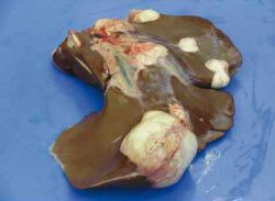
Hydatid Cysts - Donkey
| This article has been peer reviewed but is awaiting expert review. If you would like to help with this, please see more information about expert reviewing. |
Hydatid cysts - Echinococcus granulosus equinus
Introduction

Hydatid cysts and the chronic remnants of these are frequently found at post-mortems carried out at The Donkey Sanctuary. 7.8% of livers examined had a lesion indicative of a hydatid cyst. A higher incidence of hydatid cysts has been found in donkeys admitted from Ireland and Wales. In the UK, endemic areas for E. granulosus are mainly confined to the Powys area of Wales and the Outer Hebrides in Scotland. In parts of the tropics, where dogs have access to carcasses, the prevalence can be particularly high.
Hydatid cysts are the intermediate larval stage (metacestode) of the canine tapeworm, Echinococcus granulosus, which has dogs and foxes as the final host. Donkeys and horses are generally infected with the strain Echinococcus granulosus equinus, which has different host predilections from the strains that are adapted to ruminants and man. The donkey and horse act as intermediate hosts in which hydatid cysts form.
Aetiology
Donkeys ingest the eggs of E. granulosus from pastures or feed contaminated with canine faeces. Ingested tapeworm eggs migrate from the donkey’s intestine to the liver, lung and, to a lesser extent, to other organs where they form cysts and grow slowly reaching up to 6 or 7 cm diameter.
Diagnosis
Clinical signs
Most cysts are asymptomatic and are found as incidental findings at post-mortem. However, in large numbers, cysts can affect organ function. Clinical signs depend on the location of the cyst, severity of infection and size of the cyst, and are related to dysfunction of the liver.
Laboratory tests
- It is often not detected until post-mortem.
- Immunodiagnostic tests are possible, though not commonly used for diagnosis ante-mortem.
Ultrasonography
The cysts can be visualised using ultrasonography.
Treatment
Drainage of cysts can seed daughter cysts and induce anaphylaxis, although success has been reported in treating a horse with an isolated retrobulbar cyst (Summerhayes and Mantell, 1995).
Control
- Centred on eliminating the tapeworm in the final host, e.g. dogs, by regular use of a suitable dewormer such as praziquantel in endemic areas
- Dogs should be prevented from eating raw offal
- Preventing dogs and foxes from ingesting carcasses, which may harbour the cysts
- Under the Animal By-Products Order 1999, carcasses should be buried in such a way that carnivorous animals cannot gain access to them
- Prevention of faecal contamination of donkey pastures by dogs and/or their faeces should be removed from pastures
References
- Thiemann, A. (2008) Respiratory problems In Svendsen, E.D., Duncan, J. and Hadrill, D. (2008) The Professional Handbook of the Donkey, 4th edition, Whittet Books, Chapter 7
- Trawford, A. and Getachew, M. (2008) Parasites In Svendsen, E.D., Duncan, J. and Hadrill, D. (2008) The Professional Handbook of the Donkey, 4th edition, Whittet Books, Chapter 6
- Summerhayes, G.E.S., Mantell, J.A.R. (1995). ‘Ultrasonography as an aid to diagnosis and treatment of a retrolobular hydatid cyst in a horse’. Equine Veterinary Education, 7(1). pp 39-42.
References
- Thiemann, A. (2008) Respiratory problems In Svendsen, E.D., Duncan, J. and Hadrill, D. (2008) The Professional Handbook of the Donkey, 4th edition, Whittet Books, Chapter 7
|
|
This section was sponsored and content provided by THE DONKEY SANCTUARY |
|---|