Introduction
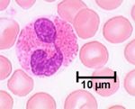
Eosinophils are a similar size to neutrophils, have a bilobed nucleus and are characterised by the large eosinophilic granules present in their cytoplasm. Produced in the bone marrow they migrate into circulation briefly before moving into tissue where they survive for around six hours. The proportion of eosinophils circulating depends on the state of the animal. Normally numbers are very low but will rise considerably during a parasitic infection or allergic reaction.
Eosinophils are mainly located in connective tissue associated with routes into the animal i.e. the respiratory, alimentary, and urogenital systems. They play key roles in reacting to parasites and allergens but have limited phagocytic ability and therefore play no role in bacterial infections.
Development
The eosinophil is a granulocyte and has a similar development to the other granulocytes; this process is called granulopoiesis. Eosinophils differ morphologically in the size and density of the cytoplasmic granules between different animal species; the granules are more prominent in the pig and the horse.
Granules
Two types of granules are present in eosinophils:
- Azurophilic granules which are present in all granulocytes and contain acid hydrolases and other enzymes.
- Specific granules contain four major proteins: Major basic protein (MBP) found in the crystalloid bodies, and then eosinophil cationic protein (ECP), eosinophil peroxidise (EPO) and eosinophil derived neurotoxin (EDN) which are all found in the granule matrix. Substances such as histaminases, cathepsins (proteases) and collagenases are also present.
Actions
Eosinophils are involved in type I hypersensitivity reactions.
In an infection or allergic reaction, eosinophil numbers are increased by the release of Il-3, Il-5 and GM-CSF by Th2 and mast cells. This causes more eosinophils to be released into the blood stream. The eosinophils are then attracted to the required site of action by chemicals known as eotaxins which are released by mast cells. Histamine and its breakdown products also act as attractants. These same products activate the eosinophil increasing its affinity to bind to IgE.
Large numbers of eosinophils in a tissue give the tissue a greenish colour.
Anti-parasitic
FC receptors on eosinophils allow them bind to parasites coated in antibodies and once bound they degranulate. The specific granules release EDN which interferes with the parasitic nervous system while MBP, ECP and EPO (EPO dissolves the carbohydrate coat of the parasite) all have anthelmintic and anti-protozoal actions. These proteins act as stimulants to mast cells causing them to release histamine and thus attracting more eosinophils. However, they also damage the animal’s tissue as well as the parasites.
Allergic
Histaminases in the specific granules breaks down histamine produced by mast cells while arylsulfatase breaks down leukotrienes. The products of these breakdowns further attracts more eosinophils.
In pathology
- See anti-parastic and allergic sections above
- Classically a cell involved in acute inflammation
- Can be quite numerous in mast cell tumours of the skin of the dog
- Eosinophilia/Eosinopenia
