Reflex Arcs - Anatomy & Physiology
Introduction
A reflex arc represents a mechanism by which a physiological function is automatically managed or regulated. Reflex arcs can be found throughout the body, ranging from skeletal muscles to smooth muscle in glands. Reflex arcs are initiated via the excitation or stimulation of specific sensory cells that are directly connected to motor neurons thus enabling motor nerve impulses to be automatically passed on to that particular muscle or gland. Therefore a basic reflex arc consists of sensory cells and their associated nerve fibers, motor nerve fibres and the ultimate muscle or gland.
Some reflex arcs can include a coordination centre within the spinal cord or brain prior to stimulation of the motor nerve. Reflex arcs can involve single or multiple segments up and down the body, although reflex arcs do not require brain input in order to function. However, the brain can act to modulate reflexes. The brain obtains its afferent information via the ascending sensory tracts of the spinal cord. The descending tracts originate from the brain to allow responses to be modulated. These tracts constitute the white matter of the spinal cord.
A number of different sensory inputs are utilised by reflex arcs, including; skin receptors, muscle spindles, the retina, the organ of Corti and the olfactory mucosa. These sensory aspects of reflex arcs feed into two main types of reflex systems in the body; autonomic reflexes and somatic reflexes.
Autonomic Reflexes
Autonomic reflexes control and regulate smooth muscle cells, cardiac muscle cells and glands. In general these reflexes contain the same basic components as somatic reflexes but a key difference is that autonomic reflexes have the ability to both stimulate or inhibit the smooth muscle/gland.
For further information regarding the basic principles of the autonomic reflex arcs and for more detailed information, please see the Autonomic Nervous System.
Somatic Reflexes
Somatic reflexes are involved in the reflex control of skeletal muscles and as such there are many different types of somatic reflexes including scratching reflexes, withdrawal reflexes and stretch reflexes and tendon reflexes. A few of these will be covered in the section below.
Tendon Reflexes
Tendons represent the weakest element of the musculoskeletal system and can be broken relatively easily compared to other aspects of the system. In some cases, muscle contractions can be so powerful that the tendon either breaks or detaches and trauma can also have a similar effect. Tendon reflexes represent a reflex arc that is designed to prevent tendon damage from occurring.
Golgi tendon organs are the sensory organs located within the tendon adjacent to the junction between the tendon and the muscle. These sensory neurons are interwoven into the collagen fibres within the tendon and are able to depolarise in response to excess changes in shape of the tendon. These golgi tendon organs are connected to inhibitory inter-neurons within the spinal cord which in turn, are connected to motor neurons innervating the muscle connected to the tendon. The result of this reflex arc is that if the sensory neurons detect tendon stretch that is excessive, the muscle will relax to reduce the load on the tendon.
Withdrawal Reflexes
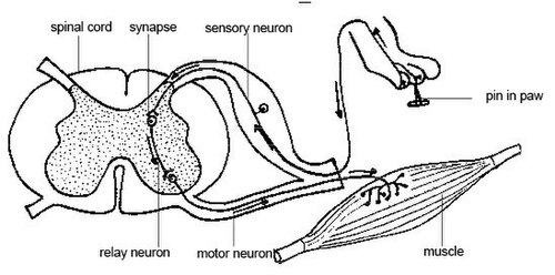
The withdrawal reflex is behind the system that automatically withdraws any area of the body that experiences pain or discomfort and is commonly used as a check for the depth of anaesthesia of surgery patients. Examples of the withdrawal reflex would be an animal that experiences heat e.g. a cat walking onto an electric hob, chemical or cold stimuli amongst many others. For the cat example, sensory neurons in the skin of the paw would be stimulated and transmit a signal to the dorsal horn of the spinal cord. Inter-neurons then connect motor neurons which innervate the muscles of the leg causing the leg muscle to flex and withdraw the paw. Muscles that would counter this movement in the leg are also inhibited to ensure the correct limb movement. In many cases the speed of this withdrawal reflex can be so fast that the paw will be retracted before the animal is consciously aware of the pain.
Stretch Reflexes
Stretch reflexes have been included here as they play an important role in posture and balance of animals and are often overlooked as this reflex functions with such efficiency it is performed totally unconsciously. Stretch reflexes are specifically used to control and coordinate the length of skeletal muscles which is particularly important in ensuring the smooth movement of limbs during locomotion.
During locomotion the body of the animal will regularly lean laterally as the limbs move. The stretch reflex ensures that during locomotion the contra-lateral muscles to the side of the lean (which will be in a stretched position) are contracted to ensure the posture of the body is brought back into a neutral position. In a similar manner to the golgi tendon organs, the stretch reflex sensory neurons are twisted around muscle spindles, called intrafusal muscle fibres. These specialised muscle fibres only constitute a small element of the muscle body. These twisted sensory neurons change shape when the muscle spindle is stretched resulting in depolarisation and the stimulation of motor neurons connected to contra-lateral antagonist muscle.
The stretch reflex is also able to activate even in muscles that are shortened as any change in muscle length will result in changes in the shape of the sensory cells. Therefore these cells provide a continuous measurement of certain muscle parameters including length and velocity of contraction. This facilitates coordination from the brain resulting in the animal being capable of very fine coordinated muscle movements that can be rapidly adjusted.
| This article has been peer reviewed but is awaiting expert review. If you would like to help with this, please see more information about expert reviewing. |
