Difference between revisions of "Abomasum - Anatomy & Physiology"
| Line 17: | Line 17: | ||
The function of the abomasum is the chemical breakdown of food. It secretes hydrochloric acid and pepsinogen. It has some intrinsic motility. Impaired motility can cause distension. The movements are slow, contractions occur first in the proximal part and are more forceful at the pyloric part. | The function of the abomasum is the chemical breakdown of food. It secretes hydrochloric acid and pepsinogen. It has some intrinsic motility. Impaired motility can cause distension. The movements are slow, contractions occur first in the proximal part and are more forceful at the pyloric part. | ||
| − | |||
==Vasculature== | ==Vasculature== | ||
The vasculature of the abomasum includes the '''cranial mesenteric artery''', the '''celiac artery''' and the '''left gastric''' and '''left gasrtoepiploic''' arteries. | The vasculature of the abomasum includes the '''cranial mesenteric artery''', the '''celiac artery''' and the '''left gastric''' and '''left gasrtoepiploic''' arteries. | ||
| − | |||
==Innervation== | ==Innervation== | ||
| Line 65: | Line 63: | ||
[[Category:Stomach - Anatomy & Physiology]] | [[Category:Stomach - Anatomy & Physiology]] | ||
| − | [[Category:To Do - AimeeHicks]] | + | [[Category:To Do - AimeeHicks]][[Category:To Do - Review]] |
Revision as of 15:57, 9 September 2010
Overview
The abomasum is the fourth chamber in the ruminant. It functions similarily to the carnivore stomach as it is glandular and functions to digest food chemically, rather than mechanically or by fermentation like the other 3 chambers of the ruminant stomach.
The abomasum differs in its position within the abdomen, depending on fullness of the other chambers of the stomach, intrinsic abomasonal activity, contractions of the rumen and reticulum (to which it is attached) and by age and pregnancy.
Displacement of the abomasum to the left or to the right is a common disorder affecting dairy cows due to high concentrate feed.
Structure
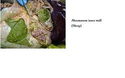
The abomasum lies upon the abdominal floor. The cranial part is split into the pylorus and body. There is also a caudal part. It is covered by the lesser omentum. It has 15-20 folds inside. The torus is at the pyloric exit. The outflow is fairly constant. There is motility at the pylorus (peristalsis). There is some control at the pyloric sphincter. The abomasum is large in newborn animals. The proximal ends of the abomasal folds forms a plug preventing reflux into the omasum. It has thin walls and a serousa covering.
Function
The function of the abomasum is the chemical breakdown of food. It secretes hydrochloric acid and pepsinogen. It has some intrinsic motility. Impaired motility can cause distension. The movements are slow, contractions occur first in the proximal part and are more forceful at the pyloric part.
Vasculature
The vasculature of the abomasum includes the cranial mesenteric artery, the celiac artery and the left gastric and left gasrtoepiploic arteries.
Innervation
The innervation of the abomasum includes the dorsal vagus nerve (CN X) and the ventral vagus nerve (CN X) (most important).
Lymphatics
Single lymph nodules are present at the junction between the epithelium and the lamina propria. Numerous small lymph nodes are scattered in the abomasal curvatures. The lymph drains to larger atrial nodes between the cardia and omasum, then to the hepatic lymph nodes.
Species Differences
Small Ruminants
The abomasum can contact the liver. The abomasum is proportionately larger than in cattle.
Links
Test yourself with the Abomasum Flashcards
Click here for Abomasum Histology
Click here for Rumen - Anatomy & Physiology
Click here for Reticulum - Anatomy & Physiology
Click here for Omasum - Anatomy & Physiology
Video links:
1.Pot 52 Lateral view of the Abdomen of a young Ruminant
2.Pot 175 Sections of the Ruminant Stomach
3.Pot 47 Ovine Omasum and Abomasum
4.Left sided topography of the Ovine Abdomen and Thorax
