Difference between revisions of "Lizard and Snake Renal Disease"
| Line 26: | Line 26: | ||
==Diagnosis== | ==Diagnosis== | ||
| − | Diagnosis of renal disease may be suspected on history and physical examination. Diagnosis includes the following: | + | Diagnosis of renal disease may be suspected on history and physical examination. Diagnosis includes the following: |
*History | *History | ||
| Line 40: | Line 40: | ||
[[Uric acid|Uric acid]] is also not a specific test for renal disease since other factors such as diet and ambient temperature also affect the levels. Increased plasma phosphate levels and a reversed [[Calcium|calcium]] to phosphate ratio are seen prior to changes in the [[Uric acid|uric acid]] level. [[lizard and Snake Imaging|Imaging]] may be useful and biopsy of the kidney gives the definitive diagnosis. | [[Uric acid|Uric acid]] is also not a specific test for renal disease since other factors such as diet and ambient temperature also affect the levels. Increased plasma phosphate levels and a reversed [[Calcium|calcium]] to phosphate ratio are seen prior to changes in the [[Uric acid|uric acid]] level. [[lizard and Snake Imaging|Imaging]] may be useful and biopsy of the kidney gives the definitive diagnosis. | ||
| − | |||
==Therapy== | ==Therapy== | ||
Revision as of 10:43, 20 August 2010
| This article has been peer reviewed but is awaiting expert review. If you would like to help with this, please see more information about expert reviewing. |
Introduction
Kidney disease in snakes and lizards is not uncommon; the main causes include gout, infectious agents and toxicoses. In an advanced state it may also be associated with metastatic soft tissue calcification, where calcium is deposited in many tissues throughout the body leading to a renal osteodystrophy. Kidney disease is common in iguanas and is a frequent cause of death in captive iguanas; most fatalities occur between 2.6 and 5.8 years, with 4.2 being the mean age with equal sex ratios.
Associated terms include: renal osteodystrophy, paradoxial metastatic calcification, metastatic mineralisation syndrome in green iguanas, and tubulonephrosis of iguanas.
The exact aetiology of renal disease in lizards is unknown but has historically been associated with:
- hypervitaminosis D or diets too high in protein, or
- visceral gout, or uric acid crystal (tophi) deposition in tissues, which may occur secondary to dehydration.
Examination
History may indicate husbandry problems such as lack of adequate water or recent treatment with possible nephrotoxic drugs. The presenting complaint is usually non-specific such as anorexia, weight loss, tremors, lethargy, flaccid paresis, distended abdomen and constipation. Physical examination may reveal nephromegaly (one or more lumps in the caudal third; in lizards, it may be palpable anterior to the pelvis or per cloaca) but there is often nothing significant found.
Diagnosis
Diagnosis of renal disease may be suspected on history and physical examination. Diagnosis includes the following:
- History
- Physical examination
- Haematology - elevated PCV if dehydrated, sometimes increased white cell count with heterophilia
- Biochemistry - inverse calcium to phosphorus ratio; elevated phosphorus; uric acid elevates late in the disease
- Radiology - poor contrast in caudal abdomen may hamper detection of nephromegaly: metastatic mineralisation seen as increased density of soft tissues, especially smooth muscle of blood vessels (most noticeable in lung fields), intestine, bladder, oviducts
- Ultrasound - kidney enlargement
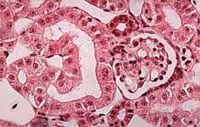
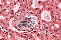
- Biopsy - kidney, confirmation of diagnosis on histology
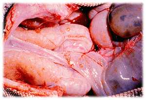
- Necropsy - when metastatic calcification is present, pulmonary, mesenteric and great vessels have a firm, gritty consistency with a thickened lumen: nephromegaly
Urea and creatinine blood levels are not useful in the diagnosis of renal disease in reptiles. Snakes are uricotelic and uric acid levels increase with kidney failure. However, it is not a sensitive test for renal disease since damage to two thirds of the kidney may be necessary before an increase will be seen.
Uric acid is also not a specific test for renal disease since other factors such as diet and ambient temperature also affect the levels. Increased plasma phosphate levels and a reversed calcium to phosphate ratio are seen prior to changes in the uric acid level. Imaging may be useful and biopsy of the kidney gives the definitive diagnosis.
Therapy
Correct diet and environmental conditions may limit the progression of the disease. Consider a low phosphorus diet. Supportive therapy is especially important since there is often no specific treatment. The most important factors include fluid therapy and being kept within the POTZ. Allopurinol and phosphate binders have been advised but are often ineffective.
Euthanasia is often the final outcome.
Prevention
As with metabolic bone disease (MBD), appropriate housing and diet will help with prevention.
Renal failure is often seen when there is some insult such as dehydration or nephrotoxic drugs. Care must be taken when drugs are prescribed to ensure that the patient is adequately hydrated. As usual good husbandry practices are essential for the optimum health of captive reptiles.


