Difference between revisions of "Protein Losing Enteropathy"
JamesSwann (talk | contribs) |
JamesSwann (talk | contribs) |
||
| Line 3: | Line 3: | ||
==Description== | ==Description== | ||
| − | '''Protein-losing enteropathy (PLE)''' refers to the loss of plasma proteins into the gastro-intestinal tract, exceeding the absorptive capacity of the intestines. PLE can be caused by: | + | '''Protein-losing enteropathy (PLE)''' refers to the loss of plasma proteins into the gastro-intestinal (GI) tract, exceeding the absorptive capacity of the intestines. PLE can be caused by: |
*Disruption to the intestinal wall due to [[Inflammation - Pathology|inflammation]] or infiltrative disease | *Disruption to the intestinal wall due to [[Inflammation - Pathology|inflammation]] or infiltrative disease | ||
| − | * | + | *Venous congestion of the GI tract |
| − | * | + | *GI [[Haemorrhage - pathology|haemorrhage]] |
| − | Hence, there are numerous causes of PLE in cats and dogs , including [[Lymphangiectasia| | + | Hence, there are numerous causes of PLE in cats and dogs , including: |
| + | *Inflammation | ||
| + | **[[Inflammatory Bowel Disease]] (including lymphocytic-plasmacytic enteritis, eosinophilic enteritis, granulomatous enteritis and histiocytic-ulcerative colitis) | ||
| + | **Uraemic gastritis and colitis | ||
| + | **[[Lymphangiectasia]] | ||
| + | **Infectious disease | ||
| + | ***Giardia duodenalis | ||
| + | ***[[Uncinaria stenocephala|Hookworm]] | ||
| + | ***[[Systemic Mycoses #Histoplasmosis|Histoplasmosis]] | ||
| + | **Chronic intussusception in juvenile animals | ||
| + | *Infiltrative disase | ||
| + | **Alimentary lymphoma | ||
| + | *Congestion | ||
| + | **Portal hypertension | ||
| + | **Posterior caval syndrome | ||
| + | **[[Heart Failure - Pathophysiology|Right-sided congestive heart failure]] | ||
| + | *GI haemorrhage | ||
| + | **e.g., hypoadrenocorticism, ischaemia | ||
| − | + | Inflammatory bowel disease and lymphoma are the most common causes of PLE in both cats and dogs, with lymphangiectasia occurring much more commonly in dogs than in cats. Chronic intussuscepta (usually occurring secondary to acute enteritis) and endoparasite infection are the most common causes of PLE in juvenile cats and dogs. | |
==Signalment== | ==Signalment== | ||
| Line 17: | Line 34: | ||
*Soft-Coated Wheaten Terrier | *Soft-Coated Wheaten Terrier | ||
**May have concurrent [[Protein Losing Nephropathy|protein-losing nephropathy]] | **May have concurrent [[Protein Losing Nephropathy|protein-losing nephropathy]] | ||
| − | |||
**Most affected animals have a common ancestor | **Most affected animals have a common ancestor | ||
| − | **Females are more commonly affected | + | **Females are more commonly affected than males |
*Yorkshire Terrier | *Yorkshire Terrier | ||
*Shar Pei | *Shar Pei | ||
| Line 33: | Line 49: | ||
==Diagnosis== | ==Diagnosis== | ||
===Clinical Signs=== | ===Clinical Signs=== | ||
| − | *Weight loss | + | *'''Weight loss''' is the most evident sign. |
| − | * | + | *'''Diarrhoea''' occurs due to the loss of protein into the GI tract and subsequent osmotic movement of fluid. Melaena may occur with GI haemorrhage. |
| − | *[[Oedema - Pathology|Oedema]], ascites and pleural effusion due to reduced plasma oncotic pressure. | + | *[[Oedema - Pathology|'''Oedema''']], '''ascites''' and '''pleural effusion''' due to reduced plasma oncotic pressure. |
| − | *Thickened intestines related to the pathological process. | + | *'''Thickened intestines''' may be detectable on abdominal palpation and this may be related to the primary pathological process. |
| − | *[[Thromboembolism|Thromboembolic]] disease due to the loss of anticoagulants such as antithrombin III. | + | *[[Thromboembolism|'''Thromboembolic]] disease''' due to the loss of anticoagulants such as antithrombin III. |
| − | *Hypocalcaemic tetany due to a reduced ability to absorb [[Vitamin D|vitamin D]]. | + | *'''Hypocalcaemic tetany''' due to a reduced ability to absorb the fat soluble [[Vitamin D|vitamin D]]. |
===Laboratory Tests=== | ===Laboratory Tests=== | ||
====Haematology==== | ====Haematology==== | ||
| − | *[[Changes in Inflammatory Cells Circulating in Blood - Pathology #Lymphopenia|Lymphopaenia]] | + | *[[Changes in Inflammatory Cells Circulating in Blood - Pathology #Lymphopenia|'''Lymphopaenia]]''' occurs with lymphangiectasia due to the loss of lymph. |
====Biochemistry==== | ====Biochemistry==== | ||
| − | *Panhypoproteinaemia | + | *'''Panhypoproteinaemia''' |
| − | **Hypoglobulinaemia with hypoalbuminaemia is a pattern more suggestive of PLE | + | **Hypoglobulinaemia with hypoalbuminaemia is a pattern more suggestive of PLE. |
| − | *Hypocholesterolaemia | + | **Albumin is usually lost in excess of globulin in protein losing nephropathy. |
| − | *[[Hypocalcaemia - Small Animal|Hypocalcaemia]] | + | *'''Hypocholesterolaemia''', especially in lymphangiectasia. |
| − | **Ionised calcium concentration should be measured to determine the significance of this finding as serum calcium concentration | + | *[[Hypocalcaemia - Small Animal|'''Hypocalcaemia''']] |
| + | **Ionised calcium concentration should be measured to determine the significance of this finding as serum calcium concentration is closely related to total protein level. | ||
====Other Tests==== | ====Other Tests==== | ||
*Measurement of faecal alpha1-protease inhibitor | *Measurement of faecal alpha1-protease inhibitor | ||
| + | **This marker has a similar molecular weight to albumin and it is lost into the GI tract in PLE. Its concentration can be measured in faeces as it is not degraded by GI enzymes. Faecal samples must be frozen on collection before submission to a laboratory in the USA. | ||
| + | *51-Chromium labelled albumin | ||
| + | **A radioactive marker (51-Chromium) is attached to recombinant albumin molecules before injection into animal. Faecal samples are collected to determine whether the labelled albumin is being lost into the GI tract. Although this test represents the 'gold standard' test, it is available only at a limited number of referral institutes. | ||
===Diagnostic Imaging=== | ===Diagnostic Imaging=== | ||
====Radiography==== | ====Radiography==== | ||
| − | *Abdominal radiographs are usually unremarkable. | + | *Abdominal radiographs |
| − | *Thoracic radiographs may show [[Effusions|pleural effusion]], metastatic neoplasia or eveidence of [[Systemic Mycoses #Histoplasmosis|histoplasmosis]]). | + | **The results are usually unremarkable but discrete mass lesions or ascites may be evident. |
| + | *Thoracic radiographs | ||
| + | **These may show the presence of [[Effusions|pleural effusion]], metastatic neoplasia or eveidence of [[Systemic Mycoses #Histoplasmosis|histoplasmosis]]). | ||
====Ultrasonography==== | ====Ultrasonography==== | ||
Revision as of 16:15, 5 July 2010
| This article is still under construction. |
Description
Protein-losing enteropathy (PLE) refers to the loss of plasma proteins into the gastro-intestinal (GI) tract, exceeding the absorptive capacity of the intestines. PLE can be caused by:
- Disruption to the intestinal wall due to inflammation or infiltrative disease
- Venous congestion of the GI tract
- GI haemorrhage
Hence, there are numerous causes of PLE in cats and dogs , including:
- Inflammation
- Inflammatory Bowel Disease (including lymphocytic-plasmacytic enteritis, eosinophilic enteritis, granulomatous enteritis and histiocytic-ulcerative colitis)
- Uraemic gastritis and colitis
- Lymphangiectasia
- Infectious disease
- Giardia duodenalis
- Hookworm
- Histoplasmosis
- Chronic intussusception in juvenile animals
- Infiltrative disase
- Alimentary lymphoma
- Congestion
- Portal hypertension
- Posterior caval syndrome
- Right-sided congestive heart failure
- GI haemorrhage
- e.g., hypoadrenocorticism, ischaemia
Inflammatory bowel disease and lymphoma are the most common causes of PLE in both cats and dogs, with lymphangiectasia occurring much more commonly in dogs than in cats. Chronic intussuscepta (usually occurring secondary to acute enteritis) and endoparasite infection are the most common causes of PLE in juvenile cats and dogs.
Signalment
The following breeds of dog may be afflicted:
- Basenji
- Lundehund
- Soft-Coated Wheaten Terrier
- May have concurrent protein-losing nephropathy
- Most affected animals have a common ancestor
- Females are more commonly affected than males
- Yorkshire Terrier
- Shar Pei
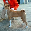 Basenji
WikiCommons
 Shar Pei
WikiCommons
 Soft Coated Wheaten Terrier
WikiCommons
 Yorkshire Terrier
WikiCommons


Diagnosis
Clinical Signs
- Weight loss is the most evident sign.
- Diarrhoea occurs due to the loss of protein into the GI tract and subsequent osmotic movement of fluid. Melaena may occur with GI haemorrhage.
- Oedema, ascites and pleural effusion due to reduced plasma oncotic pressure.
- Thickened intestines may be detectable on abdominal palpation and this may be related to the primary pathological process.
- Thromboembolic disease due to the loss of anticoagulants such as antithrombin III.
- Hypocalcaemic tetany due to a reduced ability to absorb the fat soluble vitamin D.
Laboratory Tests
Haematology
- Lymphopaenia occurs with lymphangiectasia due to the loss of lymph.
Biochemistry
- Panhypoproteinaemia
- Hypoglobulinaemia with hypoalbuminaemia is a pattern more suggestive of PLE.
- Albumin is usually lost in excess of globulin in protein losing nephropathy.
- Hypocholesterolaemia, especially in lymphangiectasia.
- Hypocalcaemia
- Ionised calcium concentration should be measured to determine the significance of this finding as serum calcium concentration is closely related to total protein level.
Other Tests
- Measurement of faecal alpha1-protease inhibitor
- This marker has a similar molecular weight to albumin and it is lost into the GI tract in PLE. Its concentration can be measured in faeces as it is not degraded by GI enzymes. Faecal samples must be frozen on collection before submission to a laboratory in the USA.
- 51-Chromium labelled albumin
- A radioactive marker (51-Chromium) is attached to recombinant albumin molecules before injection into animal. Faecal samples are collected to determine whether the labelled albumin is being lost into the GI tract. Although this test represents the 'gold standard' test, it is available only at a limited number of referral institutes.
Diagnostic Imaging
Radiography
- Abdominal radiographs
- The results are usually unremarkable but discrete mass lesions or ascites may be evident.
- Thoracic radiographs
- These may show the presence of pleural effusion, metastatic neoplasia or eveidence of histoplasmosis).
Ultrasonography
- This may reveal thickening of intestines, mesenteric lymphadenopathy or abdominal effusion.
Histopathology
- Endoscopically-guided multiple biopsies are useful. Surgical biopsy may be required for a definitive diagnosis of lymphoma and secondary lymphangiectasia. A small fatty meal could be given the night before biopsy to increase the chance of diagnosing lymphangiectasia.
- Animals with PLE have a greater risk of biopsy site dehiscence with the subsequent development of peritonitis.
Treatment
Treatment of the underlying cause of disease should be initiated, if possible.
Plasma transfusion
- This may be used to increase plasma volume. However, much of the albumin is lost in the gut and a substantial amount fails to remain in the intravascular compartment. Therefore, the extent of increase in serum albumin level is not great.
- Administration of colloid may be more suitable if it is essential to increase the plasma oncotic pressure.
Diuretics
- These can be used to reduce any ascites or pleural effusion.
- Spironolactone may be more effective than frusemide.
Antithrombotic therapy
- Treatment may be initiated with low dose aspirin to prevent the development of thrombo-embolism.
Dietary Supplementation with Calcium
- Calcium carbonate may be added to the diet if a low serum concentration of ionised calcium is documented.
Prognosis
This depends on the underlying cause.
From Pathology
Causes of PLE
- Severe inflammatory disease.
- Protein is lost in exudate.
- Lymphangiectasia.
- Loss of protein-rich lymph due to obstruction of gut lymphatics.
- Increased mucosal permeability.
- E.g. erosions, loss of tight junctions, lymphosarcoma.
- Increased loss of enterocytes (less important).
- Also:
- Immunoproliferative enteropathy
- Lymphocytic plasmacytic enteritis
- Eosinophilic enteritis
- GI ulceration/erosion
- Giardiasis
- Chronic intussusception
- Small intestinal bacterial overgrowth
- Neoplasia
- Hypoalbunimaemia causing mural oedema
- Increased activation of tissue plasminogen activator
- Systemic lupus erythematosis (SLE)
- Vascular lesion in the GI mucosa
- Chemotherapy/radiotherapy.
Pathology
- Lesions include:
- Inflammatory bowel disease
- Dilated lymphatics
- Lipogranulomatous lymphangitis.
- Intestinal crypts become dilated with mucus, sloughed epithelial cells with or without inflammatory cells.
- PLE is also associated with protein losing nephropathy (PLN).
- PLN may be a chronic sequelae to the PLE.
- Follows immune complex deposition in the glomerulus, causing glomerulonephritis or glomerulosclerosis.
- PLN causes hypoalbunaemian and hypercholesterolaemia.
- Similar PLN and PLE lesions seen in young Besenjis with immunoproliferative enteropathy and glomerulosclerosis.
References
- Ettinger, S.J. and Feldman, E. C. (2000) Textbook of Veterinary Internal Medicine Diseases of the Dog and Cat Volume 2 (Fifth Edition) W.B. Saunders Company.
- Hall, E.J, Simpson, J.W. and Williams, D.A. (2005) BSAVA Manual of Canine and Feline Gastroenterology (2nd Edition) BSAVA.
- Nelson, R.W. and Couto, C.G. (2009) Small Animal Internal Medicine (Fourth Edition) Mosby Elsevier.
- Willard, M. (2005) Protein-Losing Enteropathy in Dogs and Cats 30th World Congress of the WSAVA.