Difference between revisions of "Veterinary Education Online"

From WikiVet English
Jump to navigation Jump to search
Line 121: Line 121:
 
!
 
!
  
<h2 style="margin:0; background:#cedff2; font-size:120%; font-weight:bold; border:1px solid #a3b0bf; text-align:left; color:#000; padding:0.2em 0.4em;">Article of the Week - [[Spleen - Anatomy & Physiology|Spleen]]</h2>
+
<h2 style="margin:0; background:#cedff2; font-size:120%; font-weight:bold; border:1px solid #a3b0bf; text-align:left; color:#000; padding:0.2em 0.4em;">Article of the Week - [[Feather - Anatomy & Physiology|Feather]]</h2>
 
|-
 
|-
 
|style="color:#000;"|
 
|style="color:#000;"|

Revision as of 16:26, 3 November 2008

Welcome to WikiVet,
A collaborative initiative between the UK Vetschools to develop a comprehensive on-line veterinary knowledge base.
5,936 articles.

Content

WikiPath
Path-logo-small.png

Covers the undergraduate veterinary pathology curriculum.


WikiBugs
Bugs-logo2.png

Information about veterinary bacteriology, virology and parasitology.


Wiki Anatomy and Physiology
AntPhys Banner.png

Information about veterinary anatomy, histology and physiology.


WikiBlood
Blood-logo.png

Information about the components of blood and associated immunological diseases.


WikiClinical
Clin-logo-small.png

Clinical information.


WikiVPH
Vph-wiki.png

Information about Veterinary Public Health.


WikiDrugs
Drugs-logo2.png

Information about Pharmacology and Therapeutics.


Other resources

WikiWords

A working glossary for Wiki editors.


Feedback

An area for any comments on the content or design of the site.


e-learning Resources

Access to a range of on-line courses, videos and images.


Veterinary Education

For veterinary educators to collaborate and share expertise.


About WikiVet

More about who is involved in the WikiVet project.


WikiMap

Please try using our interactive map to find other users and add your own location.

WikiNews

23rd October 2008 - WikiDrugs Ready for Population

14th October 2008 - WikiClinical Ready for Population

All the WikiClinical stub pages are now in place. Please feel free to start populating these pages with lots of clinical knowledge. If you are having trouble editing pages please refer back to our help pages.


9th October 2008 - The Wiki is Launched
Attendees of the WikiVet Launch

The WikiVet project was officially launched at MEDEV, Newcastle University. People from varying aspects of the Veterinary, Educational, Pharmaceutical and Medical professions attended the conference. Five people also attended the virtual conference, which made for an exciting yet interactive session. We feel that the launch was a great success and look forward to watching WikiVet grow.

Article of the Week - Feather


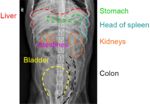
LH Canine labelled ventrodorsal abdominal organs radiograph.jpg

The spleen is a major lymphoid and blood filtration organ and is located in the left cranial abdomen. It is responsible for storing and removing erythrocytes from the blood as well as antigen surveillance of the blood and antibody production.

Development

The spleen develops in association with the digestive system in the dorsal mesogastrium, and as the stomach rotates during development the spleen comes to occupy the left cranial abdomen. Haematopoietic cells in the spleen are derived from the AGM (aorta-gonad-mesonephros) and yolk sac and as the primary lymphoid organs become established it becomes populated with T and B lymphocytes.

Structure

The spleen lies vertically on the left side of the cranial abdomen. It is attached to the greater curvature of the stomach by the gastrosplenic ligament. The spleen is covered in a enclosed in a capsule of fibrous and elastic tissue that extends into the parenchyma as trabeculae.

The parenchyma is supported by a fine mesh of reticular fibres and is divided into two types of tissue, the red and the white pulp. The red and white pulp are separated by the marginal sinus.


Click here to read more


Wiki.png
JISC.png
This project is funded by JISC and the Higher Education Academy.
HEA.png


Except where otherwise noted, content on this site is licensed under a Creative Commons Attribution 3.0 License