Coccidioidomycosis
Jump to navigation
Jump to search
- Coccidioides immitis
- Ocurs in the soil
- Respiratory infections
- Most commonly seen following dust storms
- Occurs in arid regions
- E.g. South West USA and Mexico
- Non-contageous, systemic mycosis
- Affects dogs, cattle, sheep and humans
- Mainly affects the lungs
- Dissemination can occur to other organs
- Causes nodule or granuloma formation
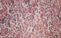
- Thick-walled spherules in tissue
- Large sporangia burst leaving 'ghost' spherules
- Saprophytic phase consists of coarse, septate, branching hyphae which fragment into thick-walled, barrel-shaped arthrospores which alternate with empty cells
- Stained by Lactose Phenol Cotton Blue
- Grows on Sabouraud's Dextrose agar and Blood agar
- Flat, moist colonies which develop a coarse, cotton-like aerial mycelium which varies from white to brown in colour
- Complement fixation test, latex agglutination and immunodiffusion tests can all be used
- A positive skin test indicates exposure
