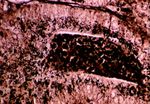
Lawsonia intracellularis
Also known as: Porcine Adenomatosis Complex — Porcine Intestinal Adenomatosis — Porcine Proliferative Enteropathy
Introduction
Porcine adenomatosis complex is a disease caused by the obligatory intracellular organism Lawsonia intracellularis, a spirochete that does not grow well in the laboratory except in enterocyte tissue culture. It is a slender, curved, microaerophilic, gram negative rod.
It is only seen in pigs, worldwide, including the United Kingdom. It is characterised by proliferative changes in the epithelium of the small and large intestinal mucosa and is excreted in small amounts in faeces of infected pigs.
Signalment
It is a disease that can affect pigs of all ages, but most severe clinical signs tend to appear in weanlings and growers. The infection is transmitted orally.
Pathogenesis
The bacterium has an affinity for porcine enterocytes. There may be synergistic relationship between L. intracellularis and intestinal flora including E. coli, Clostridium species and Bacteroides species. The intestinal organisms may produce correct oxygen tension and conditions for colonisation by L. intracellularis. Infection can only take place in presence of intestinal flora.
The disease appears in four different presentations:
Intestinal adenomatosis - The basic hyperplastic and metaplastic changes are seen in the epithelium, which causes chronic weight loss and diarrhoea.
Necrotic enteritis - Predominately affects the colon and terminal ileum and parts of the hyperplastic mucosa develop erosions and ulcerations which then become colonised by Fusiformis baceria.
Terminal ileitis - Characterised by marked hypertrophic thickening of the muscular portion of the wall of the terminal ileum and there is associated thickening of the mucosa due to hypertrophy and secondary granulomatous inflammation, which is caused by a degree of obstruction to the passage of ingesta along the bowel caused by the mucosal hypertrophy. Its appearance is very similar to Johnes disease with lots of mononuclear cells and a chronic granulomatous type of inflammation.
Proliferative haemorrhagic syndrome - The bowel shows proliferation but with ulceration and copious haemorrhage into the bowel lumen. Animals are often found dead. The pathogenesis is unclear but may involve a type of hypersensitivity reaction or secondary infection of some type.
Clinical Signs
Clinical signs are variable and can range from anything from poor weight gain to diarrhoea, weight loss, cachexia and death and appears usually in weaned pigs from age 3-4 weeks to adulthood.
It is seen often as a problem in closed, low infection herds and can lead to a decrease in production of up to 8%. It is not seen in pigs with lots of other pathogens in guts.
Diagnosis
Clinical signs of a failure to thrive and history of a closed herd plus signalment of pigs is indicative. On post mortem examination, the terminal small intestine and colon are affected by proliferation of the mucosal epithelium. Signs will include thickened mucosal epithelium, polypoid-like nodules several millimetres in diameter. There may also be undifferentiated epithelium replacing goblet cells. Its appearance is almost neoplastic.
Histologically it will appear similar to a virus induced proliferation. Organisms seen in the apical part of epithelial cells lining glands of terminal ileum, colon and caecum.
The organism from faeces or ileal mucosa can be stained by silver staining by the use of antibody in immunofluorescence or PCR or cultured in enterocyte cell lines.
Treatment and Control
Individually affected animals may be treated with tetracyclines parenterally. These animals must be isolated. In affected groups the same treatment can be given via the drinking water.
Any new genetic material into the herd should be by A.I. or embryo transfer only so as to reduce risk of introduction of the disease into the herd.
Vaccination as a control measure is now readily available and will reduce clinical signs of the disease and improve performance. Vaccine is given orally from 3 weeks of age and provides protection for up to 17 weeks. This is excellent for finishing stock, but boosters will need to be given for stock kept on as breeding herd if this is farm protocol.
References
Cowart, R.P. and Casteel, S.W. (2001) An Outline of Swine diseases: a handbook Wiley-Blackwell
Jackson, G.G. and Cockcroft, P.D. (2007) Handbook of Pig Medicine Saunders Elsevier
Straw, B.E. and Taylor, D.J. (2006) Disease of Swine Wiley-Blackwell
Taylor, D.J. (2006) Pig Diseases (Eighth edition) St Edmunsdbury Press
| This article has been peer reviewed but is awaiting expert review. If you would like to help with this, please see more information about expert reviewing. |
Error in widget FBRecommend: unable to write file /var/www/wikivet.net/extensions/Widgets/compiled_templates/wrt69a0fed5333530_44720585 Error in widget google+: unable to write file /var/www/wikivet.net/extensions/Widgets/compiled_templates/wrt69a0fed5425e46_52334664 Error in widget TwitterTweet: unable to write file /var/www/wikivet.net/extensions/Widgets/compiled_templates/wrt69a0fed54f18d4_96786197
|
| WikiVet® Introduction - Help WikiVet - Report a Problem |
