Malignant Catarrhal Fever Virus
Also know as: MCFV — MCF
Introduction
This virus is of the Herpesviridae family and the reservoir species are wildebeest and sheep, each transferring different forms of the virus. Each virus is innocuous to the reservoir host, but can sometimes cause a peculiar and fatal over T-cell division in dead end hosts. Dead end hosts include cows, kudu and some deer.
MCFV is endemic as a latent infection in Blue Wildebeest and the virus is excreted in large amounts in the periparturient period.
The disease is not all that common in UK, but is present. Outbreaks are usually sporadic and are often seen in animals mixing with sheep (carriers). In parts of Africa, you will see longer duration outbreaks, caused by different serotypes carried by Wildebeest. In deer, very serious outbreaks occur and this is probably due to infection with the sheep virus.
Signalment
Latent infection in Blue Wildebeest and sheep of all ages, breeds and sex. In dead end hosts, it can affect any sex or age, but is often seen most in youngish animals around 6 months to 1 year of age.
Clinical Signs
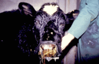
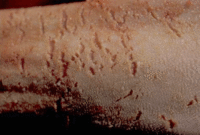
Clinical signs in cattle are severe and include necrotising lesions in the upper respiratory tract and eye: conjunctivitis and corneal oedema / opacity (keratitis or "blue eye" - characteristic feature). Pyrexia and diarrhoea with severe oculo-nasal discharge and inappetance also occurs. The animal will appear dull with ulcers on its muzzle, which exude a brown exudate. These may spread to the rest of the face and occasionally the whole body. There may also be ulcers on the tongue, dental pad, and cheeks that regularly become secondarily infected. Lymphocyte proliferation to viral antigen in the blood vessels of lymph nodes, spleen lungs, liver and kidneys progresses until death occurs. The lymph nodes eventually become completely replaced by lymphoblasts, which is a similar pathogenesis to lymphosarcoma. There may also be vasculitis with medial necrosis of blood vessels throughout body with infiltration of walls of vessels by lymphocytes.
Diagnosis
History (where and what has the animal been stocked with), signalment (deer, cattle etc) and clinical signs are quite characteristic and will lead to a preliminary diagnosis of the disease. Definitive diagnosis can be achieved by performing PCR for detection of the herpesvirus DNA.
Treatment and Control
The main control measures is to not stock Wildebeest in zoos, where possible as they are latent innocuous carries of the disease. Sheep and deer should also not be housed together.
References
Bridger, J and Russell, P (2007) Virology Study Guide, Royal Veterinary College
Divers, T.J. and Peek, S.F. (2008) Rebhun's diseases of dairy cattle Elsevier Health Scieneces
Merck & Co (2008) The Merck Veterinary Manual (Eighth Edition) Merial
Radostits, O.M, Arundel, J.H, and Gay, C.C. (2000) Veterinary Medicine: a textbook of the diseases of cattle, sheep, pigs, goats and horses Elsevier Health Sciences
| This article has been peer reviewed but is awaiting expert review. If you would like to help with this, please see more information about expert reviewing. |
Error in widget FBRecommend: unable to write file /var/www/wikivet.net/extensions/Widgets/compiled_templates/wrt69a334b89ae944_15684107 Error in widget google+: unable to write file /var/www/wikivet.net/extensions/Widgets/compiled_templates/wrt69a334b8a0a1b5_40200569 Error in widget TwitterTweet: unable to write file /var/www/wikivet.net/extensions/Widgets/compiled_templates/wrt69a334b8a4f560_96253192
|
| WikiVet® Introduction - Help WikiVet - Report a Problem |

