Periodontal Surgery - Small Animal
Introduction
Periodontal surgery is the term used for certain specific surgical techniques aimed at preserving the periodontium. Periodontal surgery techniques include closed curettage, gingivoplasty, various flap techniques, osseous surgery, guided tissue regeneration and, of course, implants. The techniques create accessibility for professional scaling and polishing, and establish a gingival morphology that facilitates plaque control by home care regimes. Some techniques are aimed at regeneration of the periodontal attachment which has been lost e.g. guided tissue regeneration.
Periodontal surgery is never a first-line treatment for periodontal disease. Conservative management of periodontal disease (i.e. thorough supra- and subgingival scaling, rootplaning and polishing), in combination with meticulous daily home care, is always the first step. If a client cannot maintain good dental hygiene for their pet, then in the interest of the well being of the animal, there is no indication for periodontal surgery.
The main objective of periodontal surgery is to contribute to the preservation of the periodontium by facilitating plaque removal and plaque control. Periodontal surgery can help achieve this by:
- Creating accessibility for professional scaling and root planing.
- Establishing a gingival morphology that facilitates plaque control by home care regimes.
- In addition, periodontal surgery may aim to regenerate a lost periodontal attachment.
Gingivoplasty
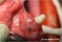
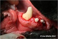
Gingivoplasty is the removal of gingival pockets by excision of the gingiva, or recontouring the gingiva to its proper anatomical form. It is indicated for the management of gingival hyperplasia. In this situation the excessive gingival tissue should be excised, leaving a normal depth of healthy gingiva.
The procedure for gingivoplasty is as follows:
- Pocket depths are measured with a graduated periodontal probe. The probe is withdrawn from the pocket and held against the outer surface of the gingiva to show the depth of the pocket and a bleeding point made. This is repeated along the circumference of the tooth.
- A beveled incision, using either a scalpel blade (No. 11 or 15) or electrosurgery is made, joining the bleeding points and recreating the scalloped edge of the normal gingival anatomy. The beveled incision is directed towards the base of the pocket or to a level slightly coronal to the apical extension of the junctional epithelium. When using electrosurgery the operator should allow for a 1 mm slough postoperatively. The electrode should be activated at the minimal effective setting in cut mode and stroked across the gingiva at the required angle. The cut surface should be pink and not bleeding if the setting is correct. Blanched tissue indicates that the setting is too high and should be reduced. To avoid overheating the tooth the electrode should not be applied to the gingiva around the same tooth for more than 5 seconds.
- The incised tissues are removed using a curette or a scaler. Remaining tissue tags are cut with a curette or a pair of scissors.
- Hemorrhage is controlled with gauze swabs and digital pressure. The crown and exposed root surfaces are then scaled and polished.
The postoperative phase is uncomfortable and analgesics are indicated for the first few days. The healing of a gingivoplasty wound is similar to that of a simple soft tissue wound. Plaque control is important to ensure adequate healing.
These animals are unlikely to accept toothbrushing immediately postoperatively, so chemical plaque control is indicated. Apply chlorhexidine gluconate gel on a piece of gauze twice daily for the first week, then once-daily in combination with toothbrushing for the second week.
Plaque control by means of daily toothbrushing and removing any predisposing causes if possible, e.g. cyclosporine administration, are necessary to prevent recurrence.
Flap Operations With or Without Osseous Surgery
Flap procedures can be used in all cases where surgical treatment of periodontal disease is indicated. These procedures can be performed with or without osseous surgery. Flap procedures are particularly useful where periodontal pockets extend beyond the mucogingival junction, the furcation is involved and/or recontouring of bony lesions is required.
The advantages of flap operations include:
- Preservation of existing keratinized gingiva.
- Increased visualization of the defect, enabling thorough debridement of dental deposits and inflamed soft tissues.
- Exposure of marginal bone, whereby the morphology of bony defects can be identified and the proper treatment carried out.
- The flap can either be replaced at its original position or shifted apically or coronally, thereby making it possible to adjust the gingival margin to the local conditions.
- The flap procedure preserves the oral epithelium.
Design of the periodontal flap used depends on the type of pocket. The most common flap used in veterinary oral surgery is usually a full flap, or one with vertical releasing incisions allowing for increased exposure, but this is considered more invasive. Another commonly used flap for periodontal surgery is the envelope flap. This is created along the arcade, without vertical incisions. Periodontal flaps are typically made full thickness, which means the periosteum is released from the underlying hard tissue and kept with the flap.
Envelope Flap: The sulcal incision is different from any other incision used in the oral cavity because the incision not only releases the gingiva from the underlying hard tissues but also excises the diseased pocket epithelium and granulation tissue. Furthermore, the sum of these incisions used to create the flap, creates a sharp marginal edge of the resulting flap, which allows for better apposition when it is replaced.
The extent of the flap should always be determined prior to any incision and all teeth in the area affected and needing treatment should be involved from the beginning to allow for a smooth incision. In most cases, carrying the incision a little mesial and distal to the target tooth is necessary for adequate visualization and access.
The full flap is created when further exposure is necessary or the flap is to be repositioned (coronally, apically, or laterally). It is a more invasive procedure with increased complication potential (e.g. hemorrhage and dehiscence) and therefore should not be performed when an envelope flap is sufficient.
| This article was written by Lisa Milella BVSc DipEVDC MRCVS. Date reviewed: 18 August 2014 |
| Endorsed by WALTHAM®, a leading authority in companion animal nutrition and wellbeing for over 50 years and the science institute for Mars Petcare. |
Error in widget FBRecommend: unable to write file /var/www/wikivet.net/extensions/Widgets/compiled_templates/wrt69a3adf97e2b39_82667253 Error in widget google+: unable to write file /var/www/wikivet.net/extensions/Widgets/compiled_templates/wrt69a3adf9844fb3_34340812 Error in widget TwitterTweet: unable to write file /var/www/wikivet.net/extensions/Widgets/compiled_templates/wrt69a3adf98a33f9_02346516
|
| WikiVet® Introduction - Help WikiVet - Report a Problem |


