Difference between revisions of "Hindbrain - Anatomy & Physiology"
| (15 intermediate revisions by 2 users not shown) | |||
| Line 1: | Line 1: | ||
| + | {{OpenPagesTop}} | ||
| + | Also known as: '''''Rhombencephalon''''' | ||
| + | |||
==Introduction== | ==Introduction== | ||
| − | The hind brain is also called the rhombencephalon and is the brain stem that provides the connection between the spinal cord and the rest of the brain | + | [[File:Brain sagittal section stem highlighted.svg|thumb|right|300px|Brain sagittal section stem highlighted]] |
| − | + | The hind brain is also called the rhombencephalon and is the brain stem that provides the connection between the spinal cord and the rest of the brain. The hind brain contains many vital structures including the '''Medulla Oblongata''', the '''Pons''' (the link between the cerebellum, forebrain and mid-brain) and the majority of the [[Cranial Nerves - Anatomy & Physiology|'''cranial nerves''']], III to XII. In general the brain stem governs essential functions that are carried out sub-consciously via reflexes. | |
| − | + | ||
| − | As well as containing numerous cranial nerves, the hind brain also contains many ' | + | As well as containing numerous cranial nerves, the hind brain also contains many ''''extra-pyramidal pathways'''' which include the '''reticular formation''', the '''olivary nucleus''' and the '''pontine nuclei'''. |
| − | + | ||
| − | + | The '''reticular formation''' is a diffuse interconnection of neurons running throughout the brain stem receiving both sensory and motor nerve tracts. This information is then passed on to higher centres in the brain such as the [[cerebrum]]. One important aspect of the reticular formation is that in order to transition from sleep to consciousness the reticular formation is required to activate the cerebral cortex. It also contains '''cerebellar pathways''' and peduncles facilitating a connection from the brain stem to the cerebellum. There are also a number of ''''pyramidal pathways'''' and afferent pathways including the cuneate and gracile pathways. | |
| − | Nuclei within the hind brain are also responsible for the reflexive control of posture and eye movement. | + | |
| + | Nuclei within the hind brain are also responsible for the '''reflexive control of posture and eye movement'''. | ||
| − | + | All elements of the hind brain are derived from the [[CNS Development - Anatomy & Physiology#Development of the Brain|developing rhombencephalon]]. | |
==Hind Brain Structures & Functions== | ==Hind Brain Structures & Functions== | ||
===Medulla Oblongata=== | ===Medulla Oblongata=== | ||
| − | The | + | The medulla oblongata can be found within the '''myelencephalon''' region of the hind-brain. Nuclei in the medulla oblongata control the level of '''heart activity''' including rate and contractility. The medulla oblongata also controls other related functions including '''blood pressure''' and distribution of blood to different organs. In conjunction with the nuclei found in the pons, the medulla oblongata also exerts an influence on '''respiratory movements'''. The respiratory centre in the medulla oblongata is affected by various types of drugs including [[opioids]] such as morphine. These types of drugs often suppress the activity of the neurones in the respiratory nuclei. |
| + | |||
===Pons=== | ===Pons=== | ||
| − | The | + | The pons can be found in the '''metencephalon''' region of the hind brain. As shown above, the pons is able to exert some influence on '''respiratory movements''' and can also influence many '''digestive processes'''. |
| + | |||
===Cranial Nerves=== | ===Cranial Nerves=== | ||
| − | + | [[Cranial Nerves - Anatomy & Physiology|Cranial nerves]] III to XII exit from the brain stem and act to innervate parts of the head, neck, viscera and the thoracic and abdominal cavities. Although most of these nerves contain both sensory and motor fibres, the sensory fibres all have their cell bodies in ganglia outside the brain stem. | |
===Cerebellum=== | ===Cerebellum=== | ||
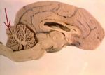
[[Image:braincerebellumarrow.jpg|thumb|right|150px|The location of the cerebellum. Image courtesy of BioMed Archive]] | [[Image:braincerebellumarrow.jpg|thumb|right|150px|The location of the cerebellum. Image courtesy of BioMed Archive]] | ||
| + | The cerebellum is located in the caudal part of the cranial cavity and is caudal to the [[Meninges - Anatomy & Physiology#Dura_mater|'tentorium cerebelli']] but dorsal to the fourth ventricle. Its gross structure is made up of a central ''''vermis'''' surrounded by two '''lateral hemispheres'''. It is attached to the brain stem via three pairs of 'peduncles'; a rostral pair to the midbrain, a middle pair to the pons and a caudal pair to the medulla oblongata. Its internal structure is composed of a cerebellar cortex which is made up of fissures on the surface that divide into lobules and then further sub divide into 'folia' or leaves. There are white matter fibres running to and from this cortex, also called '''arbor vitae'''. Within the cerebellum there are various '''nuclei''' including the '''dentate, interpositus''' and the '''fastigial'''. | ||
| − | + | The generalised function of the cerebellum is to receive information regarding any '''movement''' in progress or any intended movement via inputs from the muscles, vestibular system and motor centres of the pyramidal and extrapyramidal systems. The most important function of the cerebellum is to minimise the difference between the intended and the actual movements. The cerebellum then projects corrections regarding these movements to all motor centres of the brain via feed-back circuits between the pyramidal and extrapyramidal systems. It should be noted that the cerebellum '''cannot initiate movement'''. | |
| − | |||
| − | |||
| − | |||
| − | |||
| − | |||
| − | |||
| − | {{ | + | {{review}} |
| + | {{OpenPages}} | ||
[[Category:Nervous System - Anatomy & Physiology]] | [[Category:Nervous System - Anatomy & Physiology]] | ||
| − | [[Category: | + | [[Category:A&P Done]] |
Latest revision as of 11:45, 3 July 2012
Also known as: Rhombencephalon
Introduction
The hind brain is also called the rhombencephalon and is the brain stem that provides the connection between the spinal cord and the rest of the brain. The hind brain contains many vital structures including the Medulla Oblongata, the Pons (the link between the cerebellum, forebrain and mid-brain) and the majority of the cranial nerves, III to XII. In general the brain stem governs essential functions that are carried out sub-consciously via reflexes.
As well as containing numerous cranial nerves, the hind brain also contains many 'extra-pyramidal pathways' which include the reticular formation, the olivary nucleus and the pontine nuclei.
The reticular formation is a diffuse interconnection of neurons running throughout the brain stem receiving both sensory and motor nerve tracts. This information is then passed on to higher centres in the brain such as the cerebrum. One important aspect of the reticular formation is that in order to transition from sleep to consciousness the reticular formation is required to activate the cerebral cortex. It also contains cerebellar pathways and peduncles facilitating a connection from the brain stem to the cerebellum. There are also a number of 'pyramidal pathways' and afferent pathways including the cuneate and gracile pathways.
Nuclei within the hind brain are also responsible for the reflexive control of posture and eye movement.
All elements of the hind brain are derived from the developing rhombencephalon.
Hind Brain Structures & Functions
Medulla Oblongata
The medulla oblongata can be found within the myelencephalon region of the hind-brain. Nuclei in the medulla oblongata control the level of heart activity including rate and contractility. The medulla oblongata also controls other related functions including blood pressure and distribution of blood to different organs. In conjunction with the nuclei found in the pons, the medulla oblongata also exerts an influence on respiratory movements. The respiratory centre in the medulla oblongata is affected by various types of drugs including opioids such as morphine. These types of drugs often suppress the activity of the neurones in the respiratory nuclei.
Pons
The pons can be found in the metencephalon region of the hind brain. As shown above, the pons is able to exert some influence on respiratory movements and can also influence many digestive processes.
Cranial Nerves
Cranial nerves III to XII exit from the brain stem and act to innervate parts of the head, neck, viscera and the thoracic and abdominal cavities. Although most of these nerves contain both sensory and motor fibres, the sensory fibres all have their cell bodies in ganglia outside the brain stem.
Cerebellum
The cerebellum is located in the caudal part of the cranial cavity and is caudal to the 'tentorium cerebelli' but dorsal to the fourth ventricle. Its gross structure is made up of a central 'vermis' surrounded by two lateral hemispheres. It is attached to the brain stem via three pairs of 'peduncles'; a rostral pair to the midbrain, a middle pair to the pons and a caudal pair to the medulla oblongata. Its internal structure is composed of a cerebellar cortex which is made up of fissures on the surface that divide into lobules and then further sub divide into 'folia' or leaves. There are white matter fibres running to and from this cortex, also called arbor vitae. Within the cerebellum there are various nuclei including the dentate, interpositus and the fastigial.
The generalised function of the cerebellum is to receive information regarding any movement in progress or any intended movement via inputs from the muscles, vestibular system and motor centres of the pyramidal and extrapyramidal systems. The most important function of the cerebellum is to minimise the difference between the intended and the actual movements. The cerebellum then projects corrections regarding these movements to all motor centres of the brain via feed-back circuits between the pyramidal and extrapyramidal systems. It should be noted that the cerebellum cannot initiate movement.
| This article has been peer reviewed but is awaiting expert review. If you would like to help with this, please see more information about expert reviewing. |
Error in widget FBRecommend: unable to write file /var/www/wikivet.net/extensions/Widgets/compiled_templates/wrt6959364b3b3256_02830935 Error in widget google+: unable to write file /var/www/wikivet.net/extensions/Widgets/compiled_templates/wrt6959364b5fdce4_20357012 Error in widget TwitterTweet: unable to write file /var/www/wikivet.net/extensions/Widgets/compiled_templates/wrt6959364b7adfa9_80631199
|
| WikiVet® Introduction - Help WikiVet - Report a Problem |

