Difference between revisions of "Lizard Physical Examination"
(→Sexing) |
|||
| (25 intermediate revisions by 3 users not shown) | |||
| Line 1: | Line 1: | ||
| − | + | {{unfinished}} | |
| − | + | Consider examining in a quiet area; the use of the oculovagal reflex will allow for a more relaxed clinical examination. | |
| − | Consider examining in a quiet area; the use of the | ||
| − | |||
==Skin== | ==Skin== | ||
| − | [[Image:Lizard_dysecdysis.jpg|300px|thumb|right|'''Dysecdysis and retained skin on digits''' (Copyright © RVC)]] | + | [[Image:Lizard_dysecdysis.jpg|300px|thumb|right|'''Dysecdysis and retained skin on digits''' (Copyright © RVC and its licensors, Sean Bobbit, Sue Evans, Andrew Devare and Claire Moore. All rights reserved)]] |
| − | Examine under a bright light and with a hand lens if necessary. A common problem is retained shed especially on the digits. This may cause [[Lizard | + | Examine under a bright light and with a hand lens if necessary. A common problem is retained shed especially on the digits. This may cause [[Lizard Skin Diseases|avascular necrosis of the digits]]. |
Examine for: | Examine for: | ||
| − | * [[Lizard | + | * [[Lizard Skin Diseases|Ectoparasites]] |
| − | * Trauma due to fighting, mating or thermal [[Lizard | + | * Trauma due to fighting, mating or thermal [[Lizard Skin Diseases|burns]] |
* Discoloration (previous scars, injection sites) | * Discoloration (previous scars, injection sites) | ||
| − | * [[Lizard | + | * [[Lizard Skin Diseases|Dysecdysis]] - retained skin is often dry and brown while normal ecdysis occurring in a piecemeal fashion is flexible and transparent. Retained skin is a focus for infection and acts as a tourniquet on the tail tip and digits leading to ischaemic necrosis |
* Examine the scales for loss, scabs, blisters and haemorrhages, ensuring the ventral scales are inspected | * Examine the scales for loss, scabs, blisters and haemorrhages, ensuring the ventral scales are inspected | ||
* Assess for abnormal masses, swellings or bruised areas | * Assess for abnormal masses, swellings or bruised areas | ||
| Line 20: | Line 18: | ||
Skin folding indicates dehydration, cachexia. Assessment of dehydration in reptiles is analogous to mammals. With 5-8% dehydration there is loss of skin elasticity with a wrinkled appearance. With more severe dehydration (10-15%) the eyes are sunken and the mucous membranes become dry and sticky (consider that the oral cavity of most reptiles is relatively dry compared with mammals). | Skin folding indicates dehydration, cachexia. Assessment of dehydration in reptiles is analogous to mammals. With 5-8% dehydration there is loss of skin elasticity with a wrinkled appearance. With more severe dehydration (10-15%) the eyes are sunken and the mucous membranes become dry and sticky (consider that the oral cavity of most reptiles is relatively dry compared with mammals). | ||
| − | '''For more information on | + | '''For more information on lizard skin, see''' [[Lizard Integument|Lizard Integument]]. |
| − | |||
| − | |||
| + | '''For information on lizard skin diseases, see''' [[Lizard Skin Diseases|Lizard Skin Diseases]]. | ||
===Shedding=== | ===Shedding=== | ||
| Line 29: | Line 26: | ||
Shedding is regular and generally occurs in a piecemeal fashion. It occurs about every 4 to 6 weeks, more often for younger animals during the peak growing seasons and more slowly during winter. | Shedding is regular and generally occurs in a piecemeal fashion. It occurs about every 4 to 6 weeks, more often for younger animals during the peak growing seasons and more slowly during winter. | ||
| − | '''For | + | '''For more information on lizard shedding, see''' [[Lizard Shedding|Lizard Shedding]]. |
==Head== | ==Head== | ||
| − | [[Image:Lizard_nares.jpg|200px|thumb|right|'''Nares of an iguana''' (Copyright © RVC)]] | + | [[Image:Lizard_nares.jpg|200px|thumb|right|'''Nares of an iguana''' (Copyright © RVC and its licensors, Sean Bobbit, Sue Evans, Andrew Devare and Claire Moore. All rights reserved)]] |
| − | [[Image:Lizard_tongue.jpg|200px|thumb|right|'''The iguana tongue has a reddened split at the end of the tongue (here it is also forced to the left due to an oral granuloma)''' (Copyright © RVC)]] | + | [[Image:Lizard_eyes.jpg|200px|thumb|right|'''The eyes should be bright and clear''' (Copyright © RVC and its licensors, Sean Bobbit, Sue Evans, Andrew Devare and Claire Moore. All rights reserved)]] |
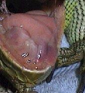
| + | [[Image:Lizard_tongue.jpg|200px|thumb|right|'''The iguana tongue has a reddened split at the end of the tongue (here it is also forced to the left due to an oral granuloma)''' (Copyright © RVC and its licensors, Sean Bobbit, Sue Evans, Andrew Devare and Claire Moore. All rights reserved)]] | ||
===Rostrum=== | ===Rostrum=== | ||
| − | * | + | * Trauma and ulcerations due to repeat escape attempts are very common in [[Water Dragon|water dragons]]. |
===Nares=== | ===Nares=== | ||
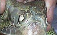
| − | * Check the nares are patent and free of discharge (some iguanids excrete salt from nasal | + | * Check the nares are patent and free of discharge (some iguanids excrete salt from nasal glands via the nares). |
===Eyes=== | ===Eyes=== | ||
| Line 46: | Line 44: | ||
* The [[Lizard Eye|eyes]] should be clear and free of discharge. | * The [[Lizard Eye|eyes]] should be clear and free of discharge. | ||
* The [[Lizard Eye|eyes]] should be bright and clear. | * The [[Lizard Eye|eyes]] should be bright and clear. | ||
| − | |||
====Ophthalmologic Examination==== | ====Ophthalmologic Examination==== | ||
| Line 55: | Line 52: | ||
* Following a general assessment of the eye and associated structures (eyelids, tear film, globe), a more detailed examination should be performed using a form of focal illumination and a magnification system. | * Following a general assessment of the eye and associated structures (eyelids, tear film, globe), a more detailed examination should be performed using a form of focal illumination and a magnification system. | ||
* Examination of the ocular fundus to assess the retina (fundoscopy) can be carried out using a direct or indirect ophthalmoscope. | * Examination of the ocular fundus to assess the retina (fundoscopy) can be carried out using a direct or indirect ophthalmoscope. | ||
| − | * [[Mydriatic|Mydriasis]] can be attempted either through general | + | * [[Mydriatic|Mydriasis]] can be attempted either through general anaesthesia or intracameral injection of neuromuscular blocking agents such as curare or d-tubocurarine. These agents can also be applied topically; however their effectiveness may be affected by the variable corneal penetration of the drugs. |
* Other diagnostic tools, such as tonometry (measurement of the intraocular pressure), stains, cytology, bacteriology, histopathology and electron microscopy, in addition to routine diagnostic tools ([[Lizard and Snake Haemotology|haemotology]], [[Lizard and Snake Biochemistry|biochemistry]], [[Lizard and Snake Imaging|radiology and ultrasound]]) can also be used to detect an ocular disease or underlying problem in lizards. | * Other diagnostic tools, such as tonometry (measurement of the intraocular pressure), stains, cytology, bacteriology, histopathology and electron microscopy, in addition to routine diagnostic tools ([[Lizard and Snake Haemotology|haemotology]], [[Lizard and Snake Biochemistry|biochemistry]], [[Lizard and Snake Imaging|radiology and ultrasound]]) can also be used to detect an ocular disease or underlying problem in lizards. | ||
| − | '''For more information on the | + | '''For more information on the eye, see''' [[Lizard Eye|Lizard Eye]]. |
| − | |||
| − | |||
===Tympanum=== | ===Tympanum=== | ||
* Check that there is no abnormal swelling. | * Check that there is no abnormal swelling. | ||
| − | '''For more information on | + | '''For more information on the lizard ear, see''' [[Lizard Ear|Lizard Ear]]. |
===Skull and mandible=== | ===Skull and mandible=== | ||
| − | * Check that there is no abnormal swelling, bony masses, soft jaw bones (esp. | + | * Check that there is no abnormal swelling, bony masses, soft jaw bones (esp. MBD). |
===Oral cavity=== | ===Oral cavity=== | ||
| Line 85: | Line 80: | ||
* Check for discharges and foreign body. | * Check for discharges and foreign body. | ||
* Observe during respiration to differentiate between discharges originating from [[Lizard Respiratory System|respiratory]] or [[Lizard Gastrointestinal System|GIT]]. | * Observe during respiration to differentiate between discharges originating from [[Lizard Respiratory System|respiratory]] or [[Lizard Gastrointestinal System|GIT]]. | ||
| − | |||
====Pharynx==== | ====Pharynx==== | ||
| Line 92: | Line 86: | ||
| − | '''For | + | '''For information on the lizard's respiratory system, see''' [[Lizard Respiratory System|Lizard Respiratory System]]. |
'''For information on the lizard's sense of taste and smell, see''' [[Lizard Taste and Smell|Lizard Taste and Smell]]. | '''For information on the lizard's sense of taste and smell, see''' [[Lizard Taste and Smell|Lizard Taste and Smell]]. | ||
==Temporomandibular joint== | ==Temporomandibular joint== | ||
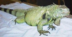
| − | [[Image:Lizard_stand.jpg|300px|thumb|right|'''In a new area, such as a consulting room, a healthy lizard will stand so that it is supporting its bodyweight (for a quick getaway if an opening appears)''' (Copyright © RVC)]] | + | [[Image:Lizard_stand.jpg|300px|thumb|right|'''In a new area, such as a consulting room, a healthy lizard will stand so that it is supporting its bodyweight (for a quick getaway if an opening appears)''' (Copyright © RVC and its licensors, Sean Bobbit, Sue Evans, Andrew Devare and Claire Moore. All rights reserved)]] |
* Check medial aspects for white deposits ([[Uric acid|uric acid]] deposits). | * Check medial aspects for white deposits ([[Uric acid|uric acid]] deposits). | ||
| Line 106: | Line 100: | ||
* Decide if swellings are bone (fibrous periosteal reactions or fractures) or soft tissue (includes abscesses). | * Decide if swellings are bone (fibrous periosteal reactions or fractures) or soft tissue (includes abscesses). | ||
* Flex and extend joints individually and assess for range of movement and pain. | * Flex and extend joints individually and assess for range of movement and pain. | ||
| − | * Check digits for | + | * Check digits for retained skin and digital necrosis. |
* Assess withdrawal reflex by pinching a digit. | * Assess withdrawal reflex by pinching a digit. | ||
Note: Muscle twitching may be associated with [[Lizard Metabolic Bone Disease|MBD]]. | Note: Muscle twitching may be associated with [[Lizard Metabolic Bone Disease|MBD]]. | ||
| + | |||
| + | '''For information on musculoskeletal problems, see''' [[Lizard Musculoskeletal Diseases|Lizard Musculoskeletal Diseases]]. | ||
==Coelomic cavity (thorax/abdomen)== | ==Coelomic cavity (thorax/abdomen)== | ||
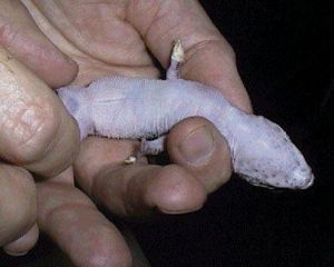
| − | [[Image:Gecko_abdominal.jpg|300px|thumb|right|'''Small lizards such as leopard geckos are delicately restrained for abdominal palpation.''' (Copyright © RVC)]] | + | [[Image:Gecko_abdominal.jpg|300px|thumb|right|'''Small lizards such as leopard geckos are delicately restrained for abdominal palpation.''' (Copyright © RVC and its licensors, Sean Bobbit, Sue Evans, Andrew Devare and Claire Moore. All rights reserved)]] |
* Requires a silent examination area for auscultation. Place stethoscope ventrally between forelimbs or laterally in axilla region for heart and lateral thorax for lungs. A thin, damp cloth between stethoscope and scales aids auscultation. [[Lizard Cardiovascular Disease|Cardiovascular problems]] are not uncommon due to calcification of bronchi and [[Lizard Cardiovascular System|major arteries]]. | * Requires a silent examination area for auscultation. Place stethoscope ventrally between forelimbs or laterally in axilla region for heart and lateral thorax for lungs. A thin, damp cloth between stethoscope and scales aids auscultation. [[Lizard Cardiovascular Disease|Cardiovascular problems]] are not uncommon due to calcification of bronchi and [[Lizard Cardiovascular System|major arteries]]. | ||
| Line 124: | Line 120: | ||
* Enlarged kidneys may be palpated by this method. Endoscopic examination should be considered. | * Enlarged kidneys may be palpated by this method. Endoscopic examination should be considered. | ||
* Examine the tail for swellings, masses, tail tip necrosis and evidence of [[Autotomy|tail loss and regeneration]]. | * Examine the tail for swellings, masses, tail tip necrosis and evidence of [[Autotomy|tail loss and regeneration]]. | ||
| − | |||
==Sexing== | ==Sexing== | ||
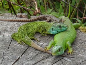
| − | [[Image:Lizard_dimorphism.jpg|300px|thumb|right|'''Sexual dimorphism in ''Lacerta bilineata'' (male: left; female: right) ''' ( | + | [[Image:Lizard_dimorphism.jpg|300px|thumb|right|'''Sexual dimorphism in ''Lacerta bilineata'' (male: left; female: right) ''' (Wikicommons)]] |
Gender identification in juvenile animals may be challenging; however, most lizard species display sexual dimorphism as adults. | Gender identification in juvenile animals may be challenging; however, most lizard species display sexual dimorphism as adults. | ||
| Line 135: | Line 130: | ||
* Sexing probes ([[Cloaca|cloacal]] probing) can also be used in iguanas and monitors but aren't as consistently accurate as they are in snakes. | * Sexing probes ([[Cloaca|cloacal]] probing) can also be used in iguanas and monitors but aren't as consistently accurate as they are in snakes. | ||
| − | * Injecting saline solution (hydrostatic eversion) into the base of the tail in order to evert the [[Hemipenes|hemipenis]] is another technique. However, care must be taken to avoid trauma to the [[Hemipenes|hemipenis]] and prevent pressure necrosis. This method can be useful for species that are difficult to sex such as [[ | + | * Injecting saline solution (hydrostatic eversion) into the base of the tail in order to evert the [[Hemipenes|hemipenis]] is another technique. However, care must be taken to avoid trauma to the [[Hemipenes|hemipenis]] and prevent pressure necrosis. This method can be useful for species that are difficult to sex such as [[Monitors|monitors]], [[Black Tegu|Tegus]], large skinks, [[Mexican Beaded Lizard|Beaded Lizards]] and [[Gila Monster|Gila Monsters]]. |
* Temporary eversion of the [[Hemipenes|hemipenes]] in anaesthetized males can be done by applying gentle pressure at the base of the tail, just caudal to the [[Cloaca|cloaca]]. | * Temporary eversion of the [[Hemipenes|hemipenes]] in anaesthetized males can be done by applying gentle pressure at the base of the tail, just caudal to the [[Cloaca|cloaca]]. | ||
| Line 150: | Line 145: | ||
==Measurement== | ==Measurement== | ||
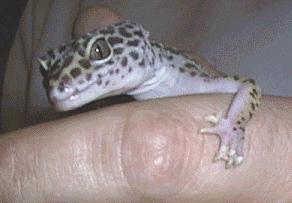
| − | Carry out normal measurement procedures, such as rostrum to | + | Carry out normal measurement procedures, such as rostrum to cloaca, cloaca to tail tip and weight. |
| − | + | [[Category:Lizard Examination|P]] | |
| − | |||
| − | |||
| − | |||
| − | |||
| − | |||
| − | |||
| − | |||
| − | |||
| − | |||
| − | |||
| − | |||
| − | [[Category:Lizard Examination| | ||
Revision as of 16:38, 20 March 2010
| This article is still under construction. |
Consider examining in a quiet area; the use of the oculovagal reflex will allow for a more relaxed clinical examination.
Skin
Examine under a bright light and with a hand lens if necessary. A common problem is retained shed especially on the digits. This may cause avascular necrosis of the digits.
Examine for:
- Ectoparasites
- Trauma due to fighting, mating or thermal burns
- Discoloration (previous scars, injection sites)
- Dysecdysis - retained skin is often dry and brown while normal ecdysis occurring in a piecemeal fashion is flexible and transparent. Retained skin is a focus for infection and acts as a tourniquet on the tail tip and digits leading to ischaemic necrosis
- Examine the scales for loss, scabs, blisters and haemorrhages, ensuring the ventral scales are inspected
- Assess for abnormal masses, swellings or bruised areas
- Assess skin elasticity
Skin folding indicates dehydration, cachexia. Assessment of dehydration in reptiles is analogous to mammals. With 5-8% dehydration there is loss of skin elasticity with a wrinkled appearance. With more severe dehydration (10-15%) the eyes are sunken and the mucous membranes become dry and sticky (consider that the oral cavity of most reptiles is relatively dry compared with mammals).
For more information on lizard skin, see Lizard Integument.
For information on lizard skin diseases, see Lizard Skin Diseases.
Shedding
Shedding is regular and generally occurs in a piecemeal fashion. It occurs about every 4 to 6 weeks, more often for younger animals during the peak growing seasons and more slowly during winter.
For more information on lizard shedding, see Lizard Shedding.
Head
Rostrum
- Trauma and ulcerations due to repeat escape attempts are very common in water dragons.
Nares
- Check the nares are patent and free of discharge (some iguanids excrete salt from nasal glands via the nares).
Eyes
Ophthalmologic Examination
The eyes are usually considered as a barometer of general health and environmental conditions; therefore a full ophthalmologic examination should be performed in all cases.
- Vision can and should be evaluated by observing certain behaviours such as a menace response.
- Following a general assessment of the eye and associated structures (eyelids, tear film, globe), a more detailed examination should be performed using a form of focal illumination and a magnification system.
- Examination of the ocular fundus to assess the retina (fundoscopy) can be carried out using a direct or indirect ophthalmoscope.
- Mydriasis can be attempted either through general anaesthesia or intracameral injection of neuromuscular blocking agents such as curare or d-tubocurarine. These agents can also be applied topically; however their effectiveness may be affected by the variable corneal penetration of the drugs.
- Other diagnostic tools, such as tonometry (measurement of the intraocular pressure), stains, cytology, bacteriology, histopathology and electron microscopy, in addition to routine diagnostic tools (haemotology, biochemistry, radiology and ultrasound) can also be used to detect an ocular disease or underlying problem in lizards.
For more information on the eye, see Lizard Eye.
Tympanum
- Check that there is no abnormal swelling.
For more information on the lizard ear, see Lizard Ear.
Skull and mandible
- Check that there is no abnormal swelling, bony masses, soft jaw bones (esp. MBD).
Oral cavity
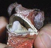
- Assess the internal extent of rostral abrasions, assess mucous membrane colour and condition (cyanosis? pallor? – normally clear and no petecchiation or oedema).
- Check for excess salivation and stomatitis.
Tongue
- Should be large and fleshy.
Glottis
- Push craniodorsally on the submandibular area to force the glottis up into better view.
- Check for discharges and foreign body.
- Observe during respiration to differentiate between discharges originating from respiratory or GIT.
Pharynx
- Examine using a bright light source.
For information on the lizard's respiratory system, see Lizard Respiratory System.
For information on the lizard's sense of taste and smell, see Lizard Taste and Smell.
Temporomandibular joint
- Check medial aspects for white deposits (uric acid deposits).
Limbs
- Assess the tone and strength of skeleton and muscle.
- Palpate for abnormal swelling or masses, especially in the joints.
- Decide if swellings are bone (fibrous periosteal reactions or fractures) or soft tissue (includes abscesses).
- Flex and extend joints individually and assess for range of movement and pain.
- Check digits for retained skin and digital necrosis.
- Assess withdrawal reflex by pinching a digit.
Note: Muscle twitching may be associated with MBD.
For information on musculoskeletal problems, see Lizard Musculoskeletal Diseases.
Coelomic cavity (thorax/abdomen)
- Requires a silent examination area for auscultation. Place stethoscope ventrally between forelimbs or laterally in axilla region for heart and lateral thorax for lungs. A thin, damp cloth between stethoscope and scales aids auscultation. Cardiovascular problems are not uncommon due to calcification of bronchi and major arteries.
- Palpate abdomen for abnormal swellings and masses (e.g. foreign bodies, cystic calculi, constipation, enlarged kidneys). Enlarged kidneys may be palpated ventrally just cranial to the pelvic inlet.
- Smaller lizards (e.g. geckos) can be transilluminated. A powerful light (e.g. endoscope) held against the body of smaller lizards or inserted into the oesophagus or cloaca of larger lizards. Care is necessary if light source produces heat. It is possible to visualise internal structure (e.g. coelomic masses).
Cloaca / tail base
- Check that the cloaca is free from faecal staining, discharge, oedema, erythema, masses or infection. The cloaca can be examined in larger species with a lubricated, gloved finger.
- Enlarged kidneys may be palpated by this method. Endoscopic examination should be considered.
- Examine the tail for swellings, masses, tail tip necrosis and evidence of tail loss and regeneration.
Sexing
Gender identification in juvenile animals may be challenging; however, most lizard species display sexual dimorphism as adults.
- The most reliable method of determining gender is usually by comparing pore sizes. Indeed, adult male lizards tend to have large femoral and precloacal pores on the ventral aspect of the thighs; on the other hand, adult females have smaller, more discrete pores, similar to those seen in juvenile male lizards.
- Sexing probes (cloacal probing) can also be used in iguanas and monitors but aren't as consistently accurate as they are in snakes.
- Injecting saline solution (hydrostatic eversion) into the base of the tail in order to evert the hemipenis is another technique. However, care must be taken to avoid trauma to the hemipenis and prevent pressure necrosis. This method can be useful for species that are difficult to sex such as monitors, Tegus, large skinks, Beaded Lizards and Gila Monsters.
- Temporary eversion of the hemipenes in anaesthetized males can be done by applying gentle pressure at the base of the tail, just caudal to the cloaca.
- In some species, such as iguanas, the hemipenes in mature males will calcify and appear visible on radiographs.
- Endoscopy can be used to visualize gonads.
- Ultrasonic evaluation of the gonads or hemipenes is also possible.
For information on the reproductive anatomy of lizards, see Lizard Reproductive System.
Measurement
Carry out normal measurement procedures, such as rostrum to cloaca, cloaca to tail tip and weight.
