Difference between revisions of "Avian Liver - Anatomy & Physiology"
Fiorecastro (talk | contribs) |
|||
| (5 intermediate revisions by 4 users not shown) | |||
| Line 1: | Line 1: | ||
| + | {{OpenPagesTop}} | ||
==Structure== | ==Structure== | ||
| − | The liver has | + | The liver has two lobes. It is dark brown coloured (except just after hatching where it is yellow). The right lobe is larger than the left lobe. It is positioned ventral and caudal to the [[Heart - Anatomy & Physiology|heart]] (as there is no diaphragm). It is closely associated to the '''[[Proventriculus - Anatomy & Physiology|proventriculus]]''' and [[Spleen - Anatomy & Physiology|spleen]]. It has a thin capsule and indistinct lobation. Two bile ducts enter the distal '''[[Duodenum - Anatomy & Physiology|duodenum]]''', one from each lobe of the liver. The duct from the right lobe is connected to the '''[[Gall Bladder - Anatomy & Physiology|gall bladder]]'''. Hepatic lobules are indistinct (except near hilus) due to a lack of '''perilobular connective tissue'''. Avian bile aids the emulsification of fats and contains amylase and lipase. |
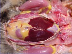
[[Image:Anatomy of the Avian Liver.jpg|thumb|right|250px|Anatomy of the Liver(Avian)- Copyright RVC 2008]] | [[Image:Anatomy of the Avian Liver.jpg|thumb|right|250px|Anatomy of the Liver(Avian)- Copyright RVC 2008]] | ||
| Line 30: | Line 31: | ||
The avian liver has '''polyhedral''' and angular cells that are larger than mammal cells. The cells have a large, spherical nucleus and the base of the cell forms the wall of the sinusoid. The cell apices communicate with the '''bile canaliculi'''. They have a granular cytoplasm. | The avian liver has '''polyhedral''' and angular cells that are larger than mammal cells. The cells have a large, spherical nucleus and the base of the cell forms the wall of the sinusoid. The cell apices communicate with the '''bile canaliculi'''. They have a granular cytoplasm. | ||
| − | '''Liver cords''' form columns around the '''interlobular bile capillary'''. The cell arrangement is simpler than in mammals. The sinusoids anastamose freely. There are ''' | + | '''Liver cords''' form columns around the '''interlobular bile capillary'''. The cell arrangement is simpler than in mammals. The sinusoids anastamose freely. There are '''Kupfer cells''' present. Fibres include, '''reticular fibres''' to support the liver cords and '''elastic fibres''' in the capsule and vessels. |
==Links== | ==Links== | ||
| − | ''' | + | '''Click here for more information on [[Liver - Anatomy & Physiology]]''' |
| − | + | {{Template:Learning | |
| + | |flashcards = [[The Avian Alimentary Tract - Anatomy & Physiology - Flashcards|Avian Alimentary Tract]] | ||
| + | |OVAM = [http://www.onlineveterinaryanatomy.net/content/interactive-avian-anatomy-liver Avian Interactive Anatomy - Liver] | ||
| + | }} | ||
| + | |||
| + | ==Webinars== | ||
| + | <rss max="10" highlight="none">https://www.thewebinarvet.com/gastroenterology-and-nutrition/webinars/feed</rss> | ||
[[Category:Avian Alimentary System - Anatomy & Physiology]] | [[Category:Avian Alimentary System - Anatomy & Physiology]] | ||
| − | [[Category: | + | [[Category:A&P Done]] |
Latest revision as of 16:15, 3 January 2023
Structure
The liver has two lobes. It is dark brown coloured (except just after hatching where it is yellow). The right lobe is larger than the left lobe. It is positioned ventral and caudal to the heart (as there is no diaphragm). It is closely associated to the proventriculus and spleen. It has a thin capsule and indistinct lobation. Two bile ducts enter the distal duodenum, one from each lobe of the liver. The duct from the right lobe is connected to the gall bladder. Hepatic lobules are indistinct (except near hilus) due to a lack of perilobular connective tissue. Avian bile aids the emulsification of fats and contains amylase and lipase.
Function
See liver function.
Vasculature
See liver vasculature.
Innervation
See liver innervation.
Lymphatics
See liver lymphatics.
Gallbladder- Species Differences
Pigeons and parrots lack a gall bladder.
Histology
The avian liver has polyhedral and angular cells that are larger than mammal cells. The cells have a large, spherical nucleus and the base of the cell forms the wall of the sinusoid. The cell apices communicate with the bile canaliculi. They have a granular cytoplasm.
Liver cords form columns around the interlobular bile capillary. The cell arrangement is simpler than in mammals. The sinusoids anastamose freely. There are Kupfer cells present. Fibres include, reticular fibres to support the liver cords and elastic fibres in the capsule and vessels.
Links
Click here for more information on Liver - Anatomy & Physiology
| Avian Liver - Anatomy & Physiology Learning Resources | |
|---|---|
 Test your knowledge using flashcard type questions |
Avian Alimentary Tract |
Anatomy Museum Resources |
Avian Interactive Anatomy - Liver |
Webinars
Failed to load RSS feed from https://www.thewebinarvet.com/gastroenterology-and-nutrition/webinars/feed: Error parsing XML for RSS

