Difference between revisions of "Snake Eye"
| (11 intermediate revisions by 3 users not shown) | |||
| Line 1: | Line 1: | ||
| − | {{ | + | {{OpenPagesTop}} |
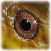
| − | Snakes are often presented to the veterinarian for eye problems so knowledge of the normal anatomy of the ophidian eye is essential. | + | [[Image:round_yellow_rat_snake_pupil.jpg|300px|thumb|right|'''Round pupil of a [[Rat snake]]''' - © RVC]] |
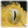
| + | [[Image:slit_palm_pit_viper_pupil.jpg|300px|thumb|right|'''Slit pupil of a [[Pit viper]]''' - © RVC]] | ||
| + | Snakes are often presented to the veterinarian for eye problems so knowledge of the normal anatomy of the ophidian eye is essential. The eyesight of snakes is relatively inefficient and the eye is reduced or absent in some fossorial snakes. They rely more heavily on the other [[Snake Special Senses|special senses]]. | ||
| − | + | '''For more information on examining snake eyes, see''' [[Snake Physical Examination]]. | |
| − | |||
| − | [[ | ||
| − | |||
| − | *'''Cornea''' - The pupil is commonly elliptical or round and may give an indication of habitat and lifestyle. Often diurnal species have round pupils and nocturnal species have vertical pupils. Coral snakes and all New World non-venomous snakes, except the boa constrictor have round pupils, while pit vipers have vertical slit pupils. | + | * '''Spectacle''' - The embryologically-fused eyelids (hence no palpebral fissure) form a transparent covering of the eye called the spectacle (also known as the brille or eyecap). Between the spectacle and the cornea are tear-like secretions. The spectacle is [[Snake Shedding|shed]] during normal [[Ecdysis|ecdysis]] but may be pathologically retained (for example if [[Snake Mites|mites]] are present, with low humidity or if [[:Category:Snake Spectacular Diseases|the eye is diseased]]). [[Snake Retained Spectacles|Retention of the spectacles]] has been associated with the development of corneal disease and panophthalmitis. |
| + | * '''Tears''' - All reptiles produce tears. In snakes, tears produce a region of lubrication between the cornea and the spectacle, allowing the free movement of the eye. Paired nasolacrimal ducts drain the sub-spectacular spaces into the mouth near the vomeronasal organ. Tears cannot overflow the eyelids, as in mammals, so if a nasolacrimal duct is damaged there is a build-up of tears under the spectacle, which leads to a [[Snake Bullous Spectaculopathy|bullous spectaculopathy]]. | ||
| + | * '''Cornea''' - The pupil is commonly elliptical or round and may give an indication of habitat and lifestyle. Often diurnal species have round pupils and nocturnal species have vertical pupils. [[coral snake|Coral snakes]] and all New World non-venomous snakes, except the [[Boa constrictor|boa constrictor]] have round pupils, while [[Pit viper|pit vipers]] have vertical slit pupils. | ||
| + | * '''Sclera''' - The eye has no ossicles (unlike other reptiles) and the sclera is composed entirely of tendinous connective tissue. | ||
| + | * '''Posterior segment''' - The retina is usually grey mottled with white or red spots and appears with semi-opaque nerve fibres radiating uniformly outwards from the optic disc that is obscured in families having a conus. It is avascular and the membrana vasculosa retinae supply nutrients. | ||
| + | ==Physiology== | ||
| + | Accommodation is similar to mammals with changes in the shape of the lens. Unlike mammals, this is under voluntary control by the action of the striated radial muscles within the ciliary body. Mammalian mydriatics therefore do not work in reptiles. However, d-tubocurarine has been used for mydriasis in reptiles. Reptiles have a rapid direct light reflex but no consensual light reflex. | ||
| − | + | {{review}} | |
| − | + | {{OpenPages}} | |
| + | |||
| + | [[Category:Snake Anatomy]] | ||
| + | [[Category:Snake_Physiology]] | ||
Latest revision as of 16:08, 18 August 2012


Snakes are often presented to the veterinarian for eye problems so knowledge of the normal anatomy of the ophidian eye is essential. The eyesight of snakes is relatively inefficient and the eye is reduced or absent in some fossorial snakes. They rely more heavily on the other special senses.
For more information on examining snake eyes, see Snake Physical Examination.
- Spectacle - The embryologically-fused eyelids (hence no palpebral fissure) form a transparent covering of the eye called the spectacle (also known as the brille or eyecap). Between the spectacle and the cornea are tear-like secretions. The spectacle is shed during normal ecdysis but may be pathologically retained (for example if mites are present, with low humidity or if the eye is diseased). Retention of the spectacles has been associated with the development of corneal disease and panophthalmitis.
- Tears - All reptiles produce tears. In snakes, tears produce a region of lubrication between the cornea and the spectacle, allowing the free movement of the eye. Paired nasolacrimal ducts drain the sub-spectacular spaces into the mouth near the vomeronasal organ. Tears cannot overflow the eyelids, as in mammals, so if a nasolacrimal duct is damaged there is a build-up of tears under the spectacle, which leads to a bullous spectaculopathy.
- Cornea - The pupil is commonly elliptical or round and may give an indication of habitat and lifestyle. Often diurnal species have round pupils and nocturnal species have vertical pupils. Coral snakes and all New World non-venomous snakes, except the boa constrictor have round pupils, while pit vipers have vertical slit pupils.
- Sclera - The eye has no ossicles (unlike other reptiles) and the sclera is composed entirely of tendinous connective tissue.
- Posterior segment - The retina is usually grey mottled with white or red spots and appears with semi-opaque nerve fibres radiating uniformly outwards from the optic disc that is obscured in families having a conus. It is avascular and the membrana vasculosa retinae supply nutrients.
Physiology
Accommodation is similar to mammals with changes in the shape of the lens. Unlike mammals, this is under voluntary control by the action of the striated radial muscles within the ciliary body. Mammalian mydriatics therefore do not work in reptiles. However, d-tubocurarine has been used for mydriasis in reptiles. Reptiles have a rapid direct light reflex but no consensual light reflex.
| This article has been peer reviewed but is awaiting expert review. If you would like to help with this, please see more information about expert reviewing. |
Error in widget FBRecommend: unable to write file /var/www/wikivet.net/extensions/Widgets/compiled_templates/wrt694a3e855ecef5_03544517 Error in widget google+: unable to write file /var/www/wikivet.net/extensions/Widgets/compiled_templates/wrt694a3e8566e988_44153812 Error in widget TwitterTweet: unable to write file /var/www/wikivet.net/extensions/Widgets/compiled_templates/wrt694a3e856d74e0_19661175
|
| WikiVet® Introduction - Help WikiVet - Report a Problem |