Difference between revisions of "Helicobacter"
| Line 1: | Line 1: | ||
| + | {{OpenPagesTop}} | ||
==Overview== | ==Overview== | ||
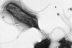
[[File:EMpylori.jpg||200px|thumb|right|''H. pylori'' - Yutaka Tsutsumi, M.D. Professor Department of Pathology Fujita Health University School of Medicine, Wikimedia Commons]] | [[File:EMpylori.jpg||200px|thumb|right|''H. pylori'' - Yutaka Tsutsumi, M.D. Professor Department of Pathology Fujita Health University School of Medicine, Wikimedia Commons]] | ||
| Line 50: | Line 51: | ||
{{review}} | {{review}} | ||
| + | |||
| + | {{OpenPages}} | ||
| + | |||
[[Category:Gastric Diseases - Dog]] | [[Category:Gastric Diseases - Dog]] | ||
Latest revision as of 18:05, 26 July 2012
Overview
Helicobacter spp. are related to Campylobacter species and Arcobacter species and are pathogens affecting the stomach. Unlike many bacterial pathogens, Helicobacter spp. are able to survive in the extremely low pH environment that exists within the stomach. The genus was first discovered in the stomach of humans in 1987.
There are several species identified in humans and many veterinary species where the incidence of some species of Helicobacter is high. Species such as H. pylori, H. felis, H. bizzozeronii, H. salomonis and H. bilis have been identified in the gastric mucosa and intestines of dogs and cats.
The genus is however considered of low pathogenic significance in veterinary species but there is possibility of zoonosis of the H. pylori species that is the causative agent of gastric disease in humans. The organism infects humans causing gastritis as well as gastric and duodenal ulcers. It has also been associated with gastric adenocarcinoma however there is no evidence for this in animals. H. pylori has not been identified in cats and is rarely seen in cats.
The major veterinary concern posed by Helicobacter spp. is the H. mustelae that has been associated with chronic gastritis and gastric ulcers in ferrets.
Characteristics
Helicobacter are gram negative rod bacteria, they can appear helical, S-shaped or curved. All species except H. canis are oxidase and catalase positive. Species of Helicobacter that colonise the gastric mucosa are urease positive as they must produce the enzyme to create a local environment with a pH of about 6-7 in order to survive.
They require enriched media, are microaerophilic and non-saccharolytic; some grow on Skirrow agar.
Clinical Infections
The significance of Helicobacter spp. in gastointestinal disease of domestic carnivores is unknown.
Up to 80% of clinically healthy dogs have Helicobacter spp. present, several species have been identified in the dog such as; H. felis, H. bizzozeronii, H. salomonis and H. bilis. Experimental infection of dogs has failed to show a consistent relationship between infection with Helicobacter and the development of pathology.
The only species known to be of veterinary importance is H. mustelae which has been associated with chronic gastritis and gastric ulcers in ferrets. Ferrets suffer from vomiting, melena, weight loss and lowered haematocrit. It has also been associated with the development of pyloric adenocarcinoma and mucosa-associated lymphoma of the stomach.
Diagnosis
Cytology from a brushing during gastric endoscopy can be performed to visualise the organism under oil immersion microscopy.
A urease test can be performed by placing gastric biopsy samples into a urease broth and observing a positive result.
Gastric biopsies can also be cultured in special media to isolate and grow Helicobacter spp.
PCR has recently been developed to demonstrate the organism in samples.
Treatment
The decision to treat for Helicobacter infections should be made on the basis of the degree of inflammation seen in the gastric mucosa of dogs in response to the organism. Treatment should lead to a resolution in inflammation and elimination of the organism.
Triple therapy using: amoxycillin and metronidazole, or tetracycline and metronidazole, combined with bismuth subsalicylate given for 2-3 weeks is usually successful in eradicating the infection. The addition of ranitidine or omeprazole can also be considered if ulcers are present.
This is also effective in ferrets infected with H. mustelae.
| Helicobacter Learning Resources | |
|---|---|
 Test your knowledge using flashcard type questions |
Small Animal Abdominal and Metabolic Disorders Q&A 15 |
References
Macpherson, C. (2000) Dog zoonoses and public health CABI
Hirsh, D. (2004) Veterinary microbiology Wiley-Blackwell
Schaer, M. (2010) Clinical medicine of the dog and cat Manson Publishing
| This article has been peer reviewed but is awaiting expert review. If you would like to help with this, please see more information about expert reviewing. |
Error in widget FBRecommend: unable to write file /var/www/wikivet.net/extensions/Widgets/compiled_templates/wrt69a20f71b8be55_26782518 Error in widget google+: unable to write file /var/www/wikivet.net/extensions/Widgets/compiled_templates/wrt69a20f71bef6e9_22211229 Error in widget TwitterTweet: unable to write file /var/www/wikivet.net/extensions/Widgets/compiled_templates/wrt69a20f71c38e81_95110676
|
| WikiVet® Introduction - Help WikiVet - Report a Problem |
