Difference between revisions of "Lizard Respiratory System"
| Line 1: | Line 1: | ||
{{review}} | {{review}} | ||
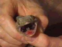
| − | [[Image:Glottis_lizard.jpg|200px|thumb|right|'''Open glottis''' (© RVC | + | [[Image:Glottis_lizard.jpg|200px|thumb|right|'''Open glottis''' (Copyright © RVC)]] |
==Lungs== | ==Lungs== | ||
Revision as of 20:38, 6 May 2010
| This article has been peer reviewed but is awaiting expert review. If you would like to help with this, please see more information about expert reviewing. |
Lungs
The trachea divides into two bronchii which open into the lungs without bronchioles. The lungs are simple hollow sacs with internal folds lined with faveoli (small sacs) for an increased surface area. In more advanced lizards, the lungs are further divided into interconnected chambers by few large septae.
- Monitors have multichambered lungs with bronchioles that each end in a faveolus.
- Chameleons have hollow, smooth-sided finger-like projections on the margins of their lungs used to inflate the body in response to predators. Some chameleons also have an accessory lung lobe projecting from the anterior trachea cranial to their forelimbs. When infected, it will fill with secretions and appear as a swelling of the ventral neck.
Respiration, which is voluntary and dependant on blood carbon dioxide pressure and temperature, is aided by expansion and contraction of the ribs as lizards lack a diaphragm.
In many lizards gas exchange occurs in the cranial part of the lung, while the caudal portion of the lung is analagous to the avian air sac.
Some lizards can revert to anaerobic metabolism during prolonged periods of apnea.
Nares
Nasal salt glands are present in herbivorous iguanid lizards such as the green iguana. These excrete excessive sodium and potassium when the plasma osmotic concentration is high. Lizards achieve this by sneezing, which expels a clear fluid that dries to a fine white powder consisting of salts. This mechanism should not be mistaken for a upper respiratory infection as it is a normal physiologic process allowing water conservation. Anterior in the roof of the mouth, the paired internal nares are a common site for discharges to accumulate and therefore a good sampling site for bacteriology when a respiratory infection is present or suspected.
Glottis
It is normally closed except during inspiration or expiration. The hard palate is reduced to allow airflow from the inner nasal opening to the glotttis. The glottis is generally quite rostral and located at the base of the tongue. This simplifies intubation and tube-feeding.
References
- Mader, D.R. (2005). Reptile Medicine and Surgery. Saunders. pp. 1264. ISBN 072169327X
