Basophils
Introduction
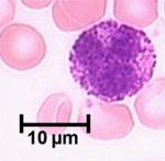
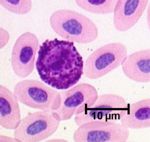
Basophils are derived from the same stem cell line as mast cells and while they are similar to mast cells, they are not identical (they are thought by some to be immature mast cells). They are the least common of all the leukocytes, are a similar size to neutrophils and eosinophils and are characterised by the large number of basophilic staining granules in their cytoplasm. They are present in the circulation but rarely found in tissue.
Development
The basophil is a granulocyte and has a similar development to the other granulocytes. This process is called granulopoiesis.
Granules
Two types of granules are present in basophils:
- Azurophilic granules which are present in all granulocytes and contain acid hydrolases and other enzymes.
- Specific granules contain heparin, histamine, leukotrienes and some lysosomes. Heparin has anti-coagulative capabilities and is also involved in assisting lipid uptake from the blood after meals. Histamine causes vasodilation.
Actions
Fc receptors on the basophil surface binds with IgE formed during allergic reactions and causes them to degranulate. The vasoactive substances released from the granules causes vasodialtion allowing for the infiltration of other leukocytes i.e. neutrophil diapedesis.
Role in pathology
- Classically a cell involved in acute inflammation
Interactions
- IgE and CD40 receptors on the basophils interact with B cells to increase IgE production
- Il-8 released by neutrophils attracts basophils
- Complement factor C3a binds to the basophil and causes degranulation

