Difference between revisions of "Musculoskeletal System Overview - Anatomy & Physiology"
| (105 intermediate revisions by 11 users not shown) | |||
| Line 1: | Line 1: | ||
| − | + | {{OpenPagesTop}} | |
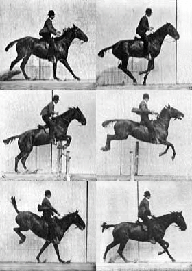
| + | [[Image:Jumping horse.gif|thumb|right|250px|'''Jumping horse''' © Eadweard Muybridge WikiMedia Commons]] | ||
| − | = | + | ==Introduction== |
| + | The musculoskeletal system includes [[Bones - Anatomy & Physiology|bones]], [[Joints - Anatomy & Physiology|joints]], [[Cartilage - Anatomy & Physiology|cartilage]], [[Muscles - Anatomy & Physiology|muscles]], ligaments and [[Muscles - Anatomy & Physiology|tendons]]. In order to describe anatomical landmarks for example for the purposes of surgery and to be able to describe different directional information, for example when recording the view of a recently taken x-ray, it is necessary to have a way of describing the [[Planes and Axes - Anatomy & Physiology|planes and axes]] that can be applied to the musculoskeletal system to pinpoint a specific anatomical area. | ||
| + | ==The Trunk== | ||
| + | The trunk consists of three segments: thorax, abdomen, and [[Pelvis - Anatomy & Physiology|pelvis]], each of which is bounded by body wall and contains a cavity. The thoracic cavity lies cranial to the diaphragm, whereas the abdominal cavity lies caudal. | ||
| − | + | Dorsally, the roof of all three cavities is formed by the [[Spinal Column - Anatomy & Physiology|spinal column]] and associated muscles. The pelvic cavity is defined by the borders of the bony pelvis and communicates with the abdominal cavity. The bony thorax includes the [[Ribs and Sternum - Anatomy & Physiology|ribs and sternum]]; the [[Ribs_and_Sternum_- Anatomy & Physiology#Thoracic_Musculature|thoracic musculature]] is predominantly associated with respiration. Knowledge of the [[Ribs_and_Sternum_- Anatomy & Physiology#Abdominal_Musculature|abdominal musculature]] is important when performing surgery on abdominal organs, and these muscles are traditionally divided into ventrolateral and sublumbar groups. | |
| − | + | ==The Head and Neck== | |
| + | The shape and size of the [[Skull and Facial Muscles - Anatomy & Physiology|skull]] varies widely, not only between species but also with age, breed and sex of similar species. The skull is divided into three components- the neurocranium, the dermatocranium and the viscerocranium. The skull also includes the [[Hyoid Apparatus - Anatomy & Physiology|hyoid apparatus]], mandible, ossicles of the middle ear and the cartilage of the [[Larynx - Anatomy & Physiology|larynx]], nose and ear. The skull protects the brain and head against injury and supports the structures of the face. In some animals the skull is also used for defensive actions, for example in horned ungulates such as red deer stags. | ||
| − | + | ===[[Pharynx - Anatomy & Physiology|Pharynx]]=== | |
| − | + | ===[[Syrinx - Anatomy & Physiology|Syrinx]]=== | |
| − | = | + | ==Limbs of the Dog, Horse and Cow== |
| − | |||
| − | + | ===[[Forelimb - Anatomy & Physiology|Forelimb]]=== | |
| + | *[[Canine Forelimb - Anatomy & Physiology|Canine Forelimb]] | ||
| − | + | *[[Bovine Forelimb - Anatomy & Physiology|Bovine Forelimb]] | |
| − | ==[[ | + | ===[[Hindlimb - Anatomy & Physiology|Hindlimb]]=== |
| − | |||
| − | |||
| − | |||
| − | |||
| − | + | *[[Canine Hindlimb - Anatomy & Physiology|Canine Hindlimb]] | |
| − | |||
| − | |||
| − | *[[ | ||
| − | + | *[[Bovine Hindlimb - Anatomy & Physiology|Bovine Hindlimb]] | |
| − | = | + | ===Phalanges=== |
| − | + | *[[Canine Phalanges - Anatomy & Physiology|Canine Phalanges]] | |
| − | + | *[[Bovine Lower Limb - Anatomy & Physiology|Bovine Lower Limb]] | |
| − | ==[[ | + | ==Topographical anatomy== |
| + | *[[Palpable Points of the Dog - Anatomy & Physiology|Palpable Points of the Dog]] | ||
| − | + | *[[Palpable Points - Horse Anatomy|Palpable Points of the Horse]] | |
| − | + | *[[Palpable Points of the Ox - Anatomy & Physiology|Palpable Points of the Ox]] | |
| − | + | {{Template:Learning | |
| − | + | |flashcards = [[:Category:Musculoskeletal System Anatomy & Physiology Flashcards|Musculoskeletal Flashcards]] | |
| − | + | |dragster= [[Canine Head and Neck Surface Anatomy Resources (I, II & III)]]<br> | |
| − | + | |OVAM = [[Musculoskeletal System Vetlogic Quiz|Musculoskeletal System Quiz]]<br>[http://www.um.es/anatvet/interactividad/ingles/alocopi/indexntscp.htm Labelled anatomy images of the canine musculoskeletal system] | |
| − | + | |Vetstream = [https://www.vetstream.com/canis/search?s=musculoskeletal+ Musculoskeletal diseases] | |
| − | + | }} | |
| − | |||
| − | |||
| − | |||
| − | |||
| − | |||
| − | |||
| − | |||
| − | |||
| − | |||
| − | |||
| − | |||
| − | |||
| − | == | ||
| − | |||
| − | |||
| − | |||
| − | |||
| + | ==References== | ||
| + | Books | ||
| + | *<div id="Dyce">{{citation|initiallast = Dyce|initialfirst = K.M|2last = Sack|2first = W.O|finallast = Wensing|finalfirst = C.J.G|year = 2002|title = Textbook of Veterinary Anatomy|ed =3rd|city = Philadelphia|pub = Saunders}}</div> | ||
| + | *O.Charnock Bradley '''The Structure of the Fowl''', 3rd ed, J.B.Lippincott Company, 1950 | ||
| + | *Konig and Liebich: '''Veterinary Anatomy of Domestic Mammals''', 3rd Edition | ||
| + | Images | ||
*''Royal Veterinary College'' Histology Department | *''Royal Veterinary College'' Histology Department | ||
| + | *''Nottingham Veterinary School'' | ||
| − | + | {{OpenPages}} | |
| − | + | [[Category:Musculoskeletal System - Anatomy & Physiology]] | |
| − | |||
| − | |||
| − | |||
| − | |||
| − | [[ | ||
Latest revision as of 20:53, 17 May 2016
Introduction
The musculoskeletal system includes bones, joints, cartilage, muscles, ligaments and tendons. In order to describe anatomical landmarks for example for the purposes of surgery and to be able to describe different directional information, for example when recording the view of a recently taken x-ray, it is necessary to have a way of describing the planes and axes that can be applied to the musculoskeletal system to pinpoint a specific anatomical area.
The Trunk
The trunk consists of three segments: thorax, abdomen, and pelvis, each of which is bounded by body wall and contains a cavity. The thoracic cavity lies cranial to the diaphragm, whereas the abdominal cavity lies caudal.
Dorsally, the roof of all three cavities is formed by the spinal column and associated muscles. The pelvic cavity is defined by the borders of the bony pelvis and communicates with the abdominal cavity. The bony thorax includes the ribs and sternum; the thoracic musculature is predominantly associated with respiration. Knowledge of the abdominal musculature is important when performing surgery on abdominal organs, and these muscles are traditionally divided into ventrolateral and sublumbar groups.
The Head and Neck
The shape and size of the skull varies widely, not only between species but also with age, breed and sex of similar species. The skull is divided into three components- the neurocranium, the dermatocranium and the viscerocranium. The skull also includes the hyoid apparatus, mandible, ossicles of the middle ear and the cartilage of the larynx, nose and ear. The skull protects the brain and head against injury and supports the structures of the face. In some animals the skull is also used for defensive actions, for example in horned ungulates such as red deer stags.
Pharynx
Syrinx
Limbs of the Dog, Horse and Cow
Forelimb
Hindlimb
Phalanges
Topographical anatomy
| Musculoskeletal System Overview - Anatomy & Physiology Learning Resources | |
|---|---|
To reach the Vetstream content, please select |
Canis, Felis, Lapis or Equis |
 Test your knowledge using drag and drop boxes |
Canine Head and Neck Surface Anatomy Resources (I, II & III) |
 Test your knowledge using flashcard type questions |
Musculoskeletal Flashcards |
Anatomy Museum Resources |
Musculoskeletal System Quiz Labelled anatomy images of the canine musculoskeletal system |
References
Books
- Dyce, K.M., Sack, W.O. and Wensing, C.J.G. (2002) Textbook of Veterinary Anatomy. 3rd ed. Philadelphia: Saunders.
- O.Charnock Bradley The Structure of the Fowl, 3rd ed, J.B.Lippincott Company, 1950
- Konig and Liebich: Veterinary Anatomy of Domestic Mammals, 3rd Edition
Images
- Royal Veterinary College Histology Department
- Nottingham Veterinary School
Error in widget FBRecommend: unable to write file /var/www/wikivet.net/extensions/Widgets/compiled_templates/wrt6633a178131041_63336467 Error in widget google+: unable to write file /var/www/wikivet.net/extensions/Widgets/compiled_templates/wrt6633a178163b31_63551111 Error in widget TwitterTweet: unable to write file /var/www/wikivet.net/extensions/Widgets/compiled_templates/wrt6633a1781915e9_86187568
|
| WikiVet® Introduction - Help WikiVet - Report a Problem |
