Difference between revisions of "Proventriculus - Anatomy & Physiology"
(→Links) |
Fiorecastro (talk | contribs) |
||
| (5 intermediate revisions by 3 users not shown) | |||
| Line 1: | Line 1: | ||
| + | |||
==Introduction== | ==Introduction== | ||
| Line 4: | Line 5: | ||
==Structure and Function== | ==Structure and Function== | ||
| − | |||
[[Image:Proventriculus Anatomy.jpg|thumb|right|250px|Proventriculus Anatomy - RVC 2008]] | [[Image:Proventriculus Anatomy.jpg|thumb|right|250px|Proventriculus Anatomy - RVC 2008]] | ||
| − | + | The proventriculus is a storage organ in fish and flesh eating birds. It is appropriate to a soft diet and secretes digestive enzymes. It contacts the left lobe of the [[Avian Liver - Anatomy & Physiology|liver]] ventrally and laterally. It is related dorso-caudally to the [[Spleen - Anatomy & Physiology|spleen]]. It is more cranial than the [[Gizzard - Anatomy & Physiology|gizzard]] and lies to the left of the midline of the bird. It is spindle/fusiform shaped. The lumen diameter is similar to the [[Oesophagus - Anatomy & Physiology|oesophagus]]. There is no oesophageal sphincter. | |
| − | The proventriculus is a storage organ in fish and flesh eating birds. It is appropriate to a soft diet and secretes digestive enzymes. It contacts the left lobe of the [[Avian Liver - Anatomy & Physiology|liver]] ventrally and laterally. It is related dorso-caudally to the [[Spleen - Anatomy & Physiology|spleen]]. It is more cranial than the [[Gizzard - Anatomy & Physiology|gizzard]] and lies to the left of the midline of the bird. It is spindle/fusiform shaped | ||
==Histology== | ==Histology== | ||
| − | |||
[[Image:Proventriculus Histology.jpg|thumb|right|250px|Proventriculus Histology - Dr. Thomas Caceci and Dr. Ihab El-Zhogby, Department of Histology, Faculty of Veterinary Medicine, Zagazig University, Egypt]] | [[Image:Proventriculus Histology.jpg|thumb|right|250px|Proventriculus Histology - Dr. Thomas Caceci and Dr. Ihab El-Zhogby, Department of Histology, Faculty of Veterinary Medicine, Zagazig University, Egypt]] | ||
| − | The proventriculus | + | The proventriculus has a '''columnar epithelium''' and the cells are basophilic. It contains '''mucous cells'''. There are '''papillae''' through which collecting ducts from glands run. '''Lamina propria''' run into the papillae. Hydrochloric acid and pepsin are produced in the glands in the submucosa. Single tubular glands are grouped into lobules with a common opening into a papilla. There is a serous membrane of '''mesothelial cells''' attached to the outer longitudinal layer of muscle. The proventriculus has 3 layers of '''lamina muscularis''' and no parietal cells. |
| − | |||
| − | |||
| − | + | {{Template:Learning | |
| + | |flashcards = [[The Avian Alimentary Tract - Anatomy & Physiology - Flashcards|Avian Alimentary Tract]] | ||
| + | |OVAM = [http://www.onlineveterinaryanatomy.net/content/interactive-avian-anatomy-digestive-system-4 Avian Interactive Anatomy - Proventriculus 1]<br>[http://www.onlineveterinaryanatomy.net/content/interactive-avian-anatomy-digestive-system-3 Avian Interactive Anatomy - Proventriculus 2] | ||
| + | }} | ||
| + | ==Webinars== | ||
| + | <rss max="10" highlight="none">https://www.thewebinarvet.com/cardiology/webinars/feed</rss> | ||
[[Category:Avian Alimentary System - Anatomy & Physiology]] | [[Category:Avian Alimentary System - Anatomy & Physiology]] | ||
| − | [[Category: | + | [[Category:A&P Done]] |
Latest revision as of 14:06, 6 January 2023
Introduction
The proventriculus is also referred to as the glandular stomach. It is connected by the isthmus to the gizzard.
Structure and Function
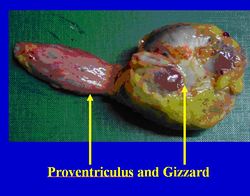
The proventriculus is a storage organ in fish and flesh eating birds. It is appropriate to a soft diet and secretes digestive enzymes. It contacts the left lobe of the liver ventrally and laterally. It is related dorso-caudally to the spleen. It is more cranial than the gizzard and lies to the left of the midline of the bird. It is spindle/fusiform shaped. The lumen diameter is similar to the oesophagus. There is no oesophageal sphincter.
Histology
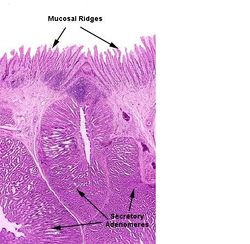
The proventriculus has a columnar epithelium and the cells are basophilic. It contains mucous cells. There are papillae through which collecting ducts from glands run. Lamina propria run into the papillae. Hydrochloric acid and pepsin are produced in the glands in the submucosa. Single tubular glands are grouped into lobules with a common opening into a papilla. There is a serous membrane of mesothelial cells attached to the outer longitudinal layer of muscle. The proventriculus has 3 layers of lamina muscularis and no parietal cells.
| Proventriculus - Anatomy & Physiology Learning Resources | |
|---|---|
 Test your knowledge using flashcard type questions |
Avian Alimentary Tract |
Anatomy Museum Resources |
Avian Interactive Anatomy - Proventriculus 1 Avian Interactive Anatomy - Proventriculus 2 |
Webinars
Failed to load RSS feed from https://www.thewebinarvet.com/cardiology/webinars/feed: Error parsing XML for RSS

