Chylous Effusion
Introduction
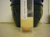
Chylous effusions are predominantly composed of chyle, the lymphatic fluid that flows through the lacteals of the small intestine and the thoracic duct in the chest. Effusions occur when the normal flow of lymph is disrupted, either by alterations in the pressure gradient between the lymphatic and systemic venous systems or by physical disruption of the lymphatic vessels. Chyle resembles milk and it is composed chiefly of fat globules (chylomicrons) with a high lymphocytic cellularity. The vast majority of chylous effusions occur in the chest (producing chylothorax) but chylous ascites may occur. The major causes of chylous effusions are:
- Reduction of the pressure gradient from the lymphatic system to the major veins
- Right-sided backward heart failure caused by cardiac tamponade, heartworm (Angiostrongylus vasorum or Dirofilaria immitis), tricuspid dysplasia or cardiomyopathy.
- Intrathoracic masses impeding venous return to the heart. Commons types of mass are thymoma and thymic lymphoma.
- Direct disruption of lymphatic flow
- Rupture of the thoracic duct due to trauma or thoracic surgery
- Erosion of lymphatics by neoplasia. The most common tumours to cause chylothorax are lymphoma, mesothelioma and tumours of the chest wall (e.g., chondrosarcomas of the ribs).
- Lung lobe torsion
- Lymphangiectasia may produce chylothorax or chylous ascites. Lymphangiectasia may be intestinal, thoracic or generalised and all forms may cause chylothorax.
- Idiopathic chylothorax may develop, especially in Afghan hounds.
Diagnosis
Clinical Signs
Effusions may occur in the thorax and, occasionally, the abdomen. This causes:
- Ascites, with which an abdominal fluid thrill will often be palpable and the abdomen may appear to be grossly swollen.
- Chylothorax causing coughing, tachypnoea and dyspnoea if severe. Dullness will be evident on thoracic percussion if a pleural effusion has developed and the heart sounds will be muffled on auscultation.
Diagnostic Imaging
Effusions are easily diagnosed by ultrasonography and this modality may also be used to guide fine needle aspiration to obtain a sample of the fluid. Effusions also produce a distinctive pattern on plain radiographs:
- With ascites, there is a loss of serosal detail due to the presence of fluid in the abdominal cavity. This appearance may also occur with large abdominal masses and in emaciated animals.
- With pleural effusions, the lung lobes are contracted and lobulation is evident. Areas of peripheral radio-opacity should be evident, especially peripherally in the chest.
Cytology
Definitive diagnosis of the type of effusion relies on collection of a sample and subsequent cytological analysis. A refractometer is frequently used to measure the specific gravity of the fluid. The following findings would be expected for a chylous effusion:
| Appearance | Opaque, milky but may be stained with blood resulting in a 'strawberry milkshake' appearance |
| Specific gravity | > 1.017 |
| Total protein | > 30g/l (variable) |
| Nucleated cells | 1.5 - 20 x 10e9/L of which the majority are small lymphocytes, mature neutrophils and variable numbers of macrophages. |
Other Tests
Pseudochyle is a type of fluid which has the same milky appearance as chyle but is actually composed of cellular debris, cholesterol micelles and lecithin globulin complexes. Pseudochyle may represent an inflammatory or neoplastic process and, although rare in animals, it may be associated with Mycobacterium infection. True chyle may be identified in the following ways:
- Chyle has a higher triglyceride concentration than plasma but a lower cholesterol concentration and it has a triglyceride: cholesterol ratio (C:T) of <1. Pseudochyle has a higher cholesterol concentration than plasma but a lower triglyceride concentration.
- When chyle is left to stand overnight, an upper 'cream' layer will become evident due to the presence of chylomicrons in the fluid, whereas pseudochyle will remain homogenous.
- The ether clearance test, in which a drop of ether was added to chyle to dissolve the lipid component, is no longer considered to be a reliable test for chylous effusions.
- Sudan III stain may be used to identify lipid droplets
Treatment
Effusions should be drained if they are causing clinical signs (of dyspnoea with chylothorax) but otherwise should be left as drainage will deplete body protein reserves. The thoracic duct can be ligated to prevent the development of chylothorax but this is not always successful and it may be difficult to identify the vessel.
A low fat diet will reduce the production of chyle as fewer fat globules are absorbed in the small intestine and lymphatic flow is reduced. This is the same rationale for the use of a low fat diet in lymphangiectasia.
Rutin is a benzopyrone drug used for the treatment of lymphoedema in humans and it may have value in the management of chylothorax, if it can be obtained. It is thought to stimulate the phagocytosis of chylomicrons by tissue macrophages.
If a tumour is found to be causing the clinical signs, resection may be attempted.
| Chylous Effusion Learning Resources | |
|---|---|
To reach the Vetstream content, please select |
Canis, Felis, Lapis or Equis |
 Test your knowledge using flashcard type questions |
Feline Medicine Q&A 11 Small Animal Emergency and Critical Care Medicine Q&A 03 |
 Search for recent publications via CAB Abstract (CABI log in required) |
Publications involving chylous effusion |
| This article has been peer reviewed but is awaiting expert review. If you would like to help with this, please see more information about expert reviewing. |
Webinars
Failed to load RSS feed from https://www.thewebinarvet.com/clinical-pathology/webinars/feed: Error parsing XML for RSS
