Difference between revisions of "Gizzard - Anatomy & Physiology"
| Line 12: | Line 12: | ||
==Histology== | ==Histology== | ||
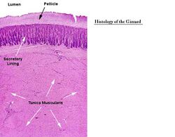
[[Image:Gizzard Histology.jpg|thumb|right|250px|Gizzard Histology- Dr. Thomas Caceci]] | [[Image:Gizzard Histology.jpg|thumb|right|250px|Gizzard Histology- Dr. Thomas Caceci]] | ||
| − | + | The gizzars has a thin, but tough '''mucous membrane'''. It has a pale, thin lining raised into ridges with 3 layers of '''lamina muscularis'''. The gizzard has cuboidal epithelium and some tubular glands present. There is a thick keratin layer to protect the muscle. | |
| − | + | The '''cuticle of koilin''' is a carbohydrate complex is present due to the solidifying of the glandular secretion. It is replenished as it is worn down. | |
| − | |||
| − | |||
| − | |||
| − | |||
| − | |||
| − | |||
| − | |||
| − | |||
| − | |||
| − | |||
==Species Differences== | ==Species Differences== | ||
Revision as of 13:00, 15 December 2010
Introduction
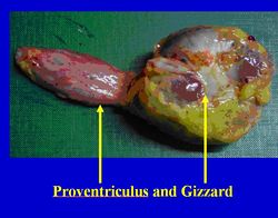
The gizzard is also referred to as the muscular stomach or ventriculus. It is connected by the isthmus to the proventriculus and to the duodenum.
Structure and Function
The gizzard allows mechanical reduction of tougher material through powerful muscular development. It is cranial to the liver and spleen (contacts the liver). It contacts the sternum and lower left abdominal wall. Dorsally the abdominal air sacs separate it from the intestines and gonads. The duodenum and pancreas lies in its caudal right surface. It is more caudal than the proventriculus and is roughly on the midline of the bird. It is lens shaped and the interior is elongated by the cranial and caudal blind sacs. The cranial blind sac contacts the proventriculus. The pylorus is on right surface next to the cranial blind sac.
The gizzard consisits of two thick masses of muscle that insert on tendonous surfaces. In seed eating birds, grit is digested to increase the grinding down of food particles. Its surface is covered by a glistening tendonous layer. The cranial and caudal extremities are formed by a powerful red muscular tissue. A circular aponeurosis is present, connecting the cranial end of the gizzard to the isthmus and the caudal end to the duodenum. It appears yellow due to bile reflux from the duodenum. When the thin muscles of the gizzard contract, food passes from the gizzard into the duodenum, when the thick muscles of the gizzard contract, food moves back into the proventriculus.
Histology
The gizzars has a thin, but tough mucous membrane. It has a pale, thin lining raised into ridges with 3 layers of lamina muscularis. The gizzard has cuboidal epithelium and some tubular glands present. There is a thick keratin layer to protect the muscle.
The cuticle of koilin is a carbohydrate complex is present due to the solidifying of the glandular secretion. It is replenished as it is worn down.
Species Differences
There is no gizzard in falconiformes (raptors etc.) or in stringiformes (owls etc.). There is also no gizzard in gulls.
Other Information
Grit should be provided in a seed eating birds diet. It is radiodense and marks out where the gizzard is located on radiographs.
Test yourself with the Avian Alimentary Tract Flashcards

