Difference between revisions of "Yeast-like fungi"
Jump to navigation
Jump to search
(Redirected page to Category:Yeast-like Fungi) |
|||
| (7 intermediate revisions by the same user not shown) | |||
| Line 1: | Line 1: | ||
| − | # | + | {{unfinished}} |
| + | |||
| + | {{toplink | ||
| + | |backcolour = | ||
| + | |linkpage =Fungi | ||
| + | |linktext =FUNGI | ||
| + | |pagetype=Bugs | ||
| + | }} | ||
| + | <br> | ||
| + | |||
| + | ==Candidosis== | ||
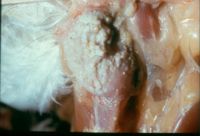
| + | [[Image:Sour Crop.jpg|thumb|right|200px|Sour Crop - Copyright Professor Andrew N. Rycroft, BSc, PHD, C. Biol.F.I.Biol., FRCPath]] | ||
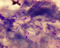
| + | [[Image:Candida.jpg|thumb|right|200px|Candida - Copyright Professor Andrew N. Rycroft, BSc, PHD, C. Biol.F.I.Biol., FRCPath]] | ||
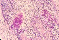
| + | [[Image:Candida in vivo.jpg|thumb|right|200px|Candida in vivo - Copyright Professor Andrew N. Rycroft, BSc, PHD, C. Biol.F.I.Biol., FRCPath]] | ||
| + | *''Candidia albicans'' is the most important species | ||
| + | **''C. tropicalis'' and ''C. pelliculosa'' are other important species | ||
| + | |||
| + | *World wide distribution | ||
| + | |||
| + | *Usually an endogenous mycoses | ||
| + | |||
| + | *Noramlly present on [[Skin - Anatomy & Physiology|skin]], [[Female Reproductive Tract -The Vagina/Vestibule - Anatomy & Physiology|vagina]] and in the [[Alimentary - Anatomy & Physiology|GI tract]] | ||
| + | |||
| + | *Immunocompromised animals may show symptoms | ||
| + | |||
| + | *Usually lesions on mucous membranes and at mucocutaneous junctions | ||
| + | |||
| + | *Many species have been implicated in bovine [[Mastitis|mastitis]] | ||
| + | |||
| + | *''C. albicans'' has been isolated in porcine stomach ulcers | ||
| + | |||
| + | *''C. rugosa'' has been implicated in pyometra in mares | ||
| + | |||
| + | *Infection of the [[Crop- Anatomy and Physiology|crop]], [[Crop- Anatomy and Physiology|oesophagus]] and [[The Avian Oral Cavity - Anatomy & Physiology|mouth]] occur in poultry and other birds leading to '''sour crop''' | ||
| + | **White-grey lesions in mouth which adhere loosly to the mucous membrane | ||
| + | **[[Crop- Anatomy and Physiology|Crop]] wall may be thickened | ||
| + | **[[Crop- Anatomy and Physiology|Crop]] wall may be covered by a yellow-white necrotic material | ||
| + | **Underlying tissue is inflammed | ||
| + | |||
| + | *Causes thrush in humans | ||
| + | |||
| + | *''C. albicans'' causes metritis and vaginitis in mares and genital candidiosis in stallions (and bulls) | ||
| + | |||
| + | *Skin scrapings in 20% KOH for microscopy | ||
| + | |||
| + | *Diphtheritic membranes, pus and fluids can be examined by Lactophenol Cotton Blue and stained by Gram or Methylene Blue stain | ||
| + | |||
| + | *Gram positive, oval, thin-walled budding cells with hyphal fragments | ||
| + | |||
| + | *Grow on Blood agar and Sabouraud's Dextrose agar producing soft, creamy colonies in 24-48 hours | ||
| + | |||
| + | *Grossly: | ||
| + | **Exudative, papular, pustular to ulcerative dermatitis | ||
| + | **Stomatitis and otitis externa may develop | ||
| + | |||
| + | *Microscopically: | ||
| + | **Spongiotic neutrophilic pustular inflammation | ||
| + | **Parakeratosis | ||
| + | **Ulcerations | ||
| + | **Superficial exudate containing organisms | ||
| + | |||
| + | *''Candida'' spp. in [[Mycotic skin infections - Pathology#Candidiasis|candidiasis]] | ||
| + | |||
| + | ==Cryptococcosis== | ||
| + | |||
| + | *Over 19 species | ||
| + | **''C. neoformans'' only major pathogen | ||
| + | |||
| + | *Worldwide | ||
| + | |||
| + | *Occurs in high concentrations in pigeon droppings (high creatinine concentration) | ||
| + | **The pigeon is not infected | ||
| + | **''C. neoformis'' colonise the droppings after they have been excreted | ||
| + | **Also found in fruit, milk and soil | ||
| + | |||
| + | *Exogenous, inhaled infection which is generally sporadic (non-contageous) | ||
| + | **Can also be absorbed via skin penetration and ingestion | ||
| + | |||
| + | *May be a primary pathogen or opportunistic | ||
| + | |||
| + | *Targets the [[Cardiorespiratory System - Anatomy & Physiology|respiratory system]] | ||
| + | **Including the [[Paranasal sinuses - Anatomy & Physiology|paranasal sinuses]] | ||
| + | **Also can be systemic, cutaneous, visceral, skeletal or ocular | ||
| + | |||
| + | *Causes sporadic mastitis in cattle | ||
| + | **Can spread within the herd | ||
| + | |||
| + | *Affects the [[Nervous and Special Senses - Anatomy & Physiology#Central Nervous System (CNS)|CNS]] of dogs and cats | ||
| + | **[[Paranasal sinuses - Anatomy & Physiology|paranasal sinuses]] and [[Pharynx - Anatomy & Physiology|pharynx]] can be infected with dissemination to the [[Nervous and Special Senses - Anatomy & Physiology#Central Nervous System (CNS)|CNS]] and other tissues | ||
| + | ***E.g. [[Lungs - Anatomy & Physiology|Lungs]], [[Urinary System - Anatomy & Physiology#The Kidney|kidneys]] and [[Joints - Anatomy & Physiology|joints]] | ||
| + | **Also causes subcutaneous granulomas | ||
| + | **The tip of the nose is a common site of infection in cats | ||
| + | ***See [[Respiratory Fungal Infections - Pathology#In Cats|here]] | ||
| + | |||
| + | *Causes myxoma-like lesions of the [[Lungs - Anatomy & Physiology|lung]] and [[Lips - Anatomy & Physiology|lip]] in horses | ||
| + | |||
| + | *Causes cryptococcal meningitis in humans | ||
| + | |||
| + | *Also affects dolphins, foxes, ferrets, monkeys, birds, cheetahs and guinea-pigs | ||
| + | |||
| + | *Large yeast with capsule seen using India ink stain | ||
| + | |||
| + | *Stains with PAS (Periodic acis Schiff) | ||
| + | |||
| + | *Gram positive | ||
| + | |||
| + | *Grows on blood agar and Sabouraud's Dextrose agar forming white, granular colonies which become slimy, mucoid and turn creamy/brown within a week | ||
| + | |||
| + | *Species identified by carbohydrate assimilation tests | ||
| + | |||
| + | *Antigen and antibody should be tested for as [[Immunoglobulins - WikiBlood|antibody]] formed by the body is soon overwhelmed and neutralised by abundent polysaccharide antigen from the capsule in active, systemic infections | ||
| + | **Latex agglutination for [[Adaptive Immune System - WikiBlood#Actions of the Adaptive Immune System|antigen]], complement fixation, ELISA and IFAT can be used | ||
| + | |||
| + | ==Geotrichosis== | ||
| + | |||
| + | *''G. candidum'' | ||
| + | |||
| + | *Rare | ||
| + | |||
| + | *Two forms: the yeast-like (glaborous) and fluffy | ||
| + | |||
| + | *Affects a wide range of species | ||
| + | |||
| + | *Usually diagnosed post-mortem | ||
| + | |||
| + | *Affects the mucous membranes, udder, [[Bronchi and bronchioles - Anatomy & Physiology|bronchi]] and [[Lungs - Anatomy & Physiology|lungs]] | ||
| + | |||
| + | *Usually mild, causing suppurative granulomas | ||
| + | |||
| + | *Can be recovered from otitis externa infections in dogs | ||
| + | |||
| + | *Organisms appear as rectangular or spherical arthrospores on wet mounts | ||
| + | **Thick walled, non-budding, gram positive | ||
| + | |||
| + | *Grow on Sabouraud's Dextrose agar | ||
| + | **Membranous colonies | ||
| + | **Do not grow well on blood agar | ||
| + | |||
| + | ==''Malassezia pachydermidis''== | ||
| + | [[Image:Malassezia pachydermidis.jpg|thumb|right|150px|''Malassezia pachydermidis'' - Copyright Professor Andrew N. Rycroft, BSc, PHD, C. Biol.F.I.Biol., FRCPath]] | ||
| + | *Normally present in oily areas on the external [[Ear - Anatomy & Physiology|ear]] canal and [[Skin - Anatomy & Physiology|skin]] in dogs | ||
| + | **Some strains have been recovered from the [[Ear - Anatomy & Physiology|ear]] canal of cats | ||
| + | |||
| + | *Bottle-shaped, small budding cells, non-mycelial | ||
| + | |||
| + | *Gram stain shows purple yeast cells with a very wide base | ||
| + | |||
| + | *Grows on Sabouraud's Dextrose agar in 2 weeks of incubation at room temperature | ||
| + | |||
| + | *Greenish discolouration on blood agar | ||
| + | |||
| + | *Clinical disease may cause yeast to proliferate and cause infection | ||
| + | |||
| + | *Grossly: | ||
| + | **Regional lesions: muzzle, [[Ear - Anatomy & Physiology|ears]], interdigital, perianal | ||
| + | **Or generalised disease | ||
| + | **Erythematous, hyperpigmented, [[Skin Glossary - Pathology|lichenified]] and scaly lesions with alopecia | ||
| + | |||
| + | *Microscopically: | ||
| + | **[[Skin Glossary - Pathology|hyperkeratosis, parakeratosis]] | ||
| + | **Spongiotic pustular dermatitis | ||
| + | **[[Skin Glossary - Pathology|Acanthosis]] | ||
| + | **Organisms are usually present- minimum 3-5 yeasts per high-power field must be found to imply cause of disease | ||
| + | |||
| + | *''Malassezia pachydermis'' in [[Mycotic skin infections - Pathology#Malassezia dermatitis|malassezia dermatitis]] | ||
| + | |||
| + | ==''Rhodotorula''== | ||
| + | |||
| + | *''Rhodotorula minuta'' and ''R. rubra'' | ||
| + | |||
| + | *Canine [[Ear - Anatomy & Physiology|ear]] infections | ||
| + | |||
| + | *Equine [[Female Reproductive Tract -The Uterus - Anatomy & Physiology|uterus]] | ||
| + | |||
| + | *Seldom seen in animal infections | ||
| + | |||
| + | ==''Torulopsis glabrata''== | ||
| + | |||
| + | *Commensal in animals and is found in the soil | ||
| + | |||
| + | *Implicated in cases of: | ||
| + | **Pyelonephritis, pneumonia, septicaemia and meningitis (humans) | ||
| + | **Mastitis and abortion in cattle | ||
| + | **Systemic infection of monkeys and dogs | ||
| + | |||
| + | ==Trichosporonosis== | ||
| + | |||
| + | *Found in soil | ||
| + | |||
| + | *Deuteromycetes yeast | ||
| + | |||
| + | *''Trichosporonosis beigelii'' | ||
| + | **Implicated in feline nasal granuloma, [[Mycotic skin infections - Pathology#Superficial mycoses|skin infections]] in horses and monkeys, mastitis in cattle and sheep and in feline bladder infections | ||
| + | |||
| + | *''T. capitum'' implicated in bovine mastitis | ||
| + | |||
| + | ==Further Links== | ||
| + | |||
| + | *[[Mycotic skin infections - Pathology]] | ||
| + | |||
| + | *[[Antifungal Drugs]] | ||
Revision as of 16:04, 14 August 2009
| This article is still under construction. |
|
|
Candidosis
- Candidia albicans is the most important species
- C. tropicalis and C. pelliculosa are other important species
- World wide distribution
- Usually an endogenous mycoses
- Immunocompromised animals may show symptoms
- Usually lesions on mucous membranes and at mucocutaneous junctions
- Many species have been implicated in bovine mastitis
- C. albicans has been isolated in porcine stomach ulcers
- C. rugosa has been implicated in pyometra in mares
- Infection of the crop, oesophagus and mouth occur in poultry and other birds leading to sour crop
- Causes thrush in humans
- C. albicans causes metritis and vaginitis in mares and genital candidiosis in stallions (and bulls)
- Skin scrapings in 20% KOH for microscopy
- Diphtheritic membranes, pus and fluids can be examined by Lactophenol Cotton Blue and stained by Gram or Methylene Blue stain
- Gram positive, oval, thin-walled budding cells with hyphal fragments
- Grow on Blood agar and Sabouraud's Dextrose agar producing soft, creamy colonies in 24-48 hours
- Grossly:
- Exudative, papular, pustular to ulcerative dermatitis
- Stomatitis and otitis externa may develop
- Microscopically:
- Spongiotic neutrophilic pustular inflammation
- Parakeratosis
- Ulcerations
- Superficial exudate containing organisms
- Candida spp. in candidiasis
Cryptococcosis
- Over 19 species
- C. neoformans only major pathogen
- Worldwide
- Occurs in high concentrations in pigeon droppings (high creatinine concentration)
- The pigeon is not infected
- C. neoformis colonise the droppings after they have been excreted
- Also found in fruit, milk and soil
- Exogenous, inhaled infection which is generally sporadic (non-contageous)
- Can also be absorbed via skin penetration and ingestion
- May be a primary pathogen or opportunistic
- Targets the respiratory system
- Including the paranasal sinuses
- Also can be systemic, cutaneous, visceral, skeletal or ocular
- Causes sporadic mastitis in cattle
- Can spread within the herd
- Affects the CNS of dogs and cats
- Causes cryptococcal meningitis in humans
- Also affects dolphins, foxes, ferrets, monkeys, birds, cheetahs and guinea-pigs
- Large yeast with capsule seen using India ink stain
- Stains with PAS (Periodic acis Schiff)
- Gram positive
- Grows on blood agar and Sabouraud's Dextrose agar forming white, granular colonies which become slimy, mucoid and turn creamy/brown within a week
- Species identified by carbohydrate assimilation tests
- Antigen and antibody should be tested for as antibody formed by the body is soon overwhelmed and neutralised by abundent polysaccharide antigen from the capsule in active, systemic infections
- Latex agglutination for antigen, complement fixation, ELISA and IFAT can be used
Geotrichosis
- G. candidum
- Rare
- Two forms: the yeast-like (glaborous) and fluffy
- Affects a wide range of species
- Usually diagnosed post-mortem
- Usually mild, causing suppurative granulomas
- Can be recovered from otitis externa infections in dogs
- Organisms appear as rectangular or spherical arthrospores on wet mounts
- Thick walled, non-budding, gram positive
- Grow on Sabouraud's Dextrose agar
- Membranous colonies
- Do not grow well on blood agar
Malassezia pachydermidis
- Normally present in oily areas on the external ear canal and skin in dogs
- Some strains have been recovered from the ear canal of cats
- Bottle-shaped, small budding cells, non-mycelial
- Gram stain shows purple yeast cells with a very wide base
- Grows on Sabouraud's Dextrose agar in 2 weeks of incubation at room temperature
- Greenish discolouration on blood agar
- Clinical disease may cause yeast to proliferate and cause infection
- Grossly:
- Regional lesions: muzzle, ears, interdigital, perianal
- Or generalised disease
- Erythematous, hyperpigmented, lichenified and scaly lesions with alopecia
- Microscopically:
- hyperkeratosis, parakeratosis
- Spongiotic pustular dermatitis
- Acanthosis
- Organisms are usually present- minimum 3-5 yeasts per high-power field must be found to imply cause of disease
- Malassezia pachydermis in malassezia dermatitis
Rhodotorula
- Rhodotorula minuta and R. rubra
- Canine ear infections
- Equine uterus
- Seldom seen in animal infections
Torulopsis glabrata
- Commensal in animals and is found in the soil
- Implicated in cases of:
- Pyelonephritis, pneumonia, septicaemia and meningitis (humans)
- Mastitis and abortion in cattle
- Systemic infection of monkeys and dogs
Trichosporonosis
- Found in soil
- Deuteromycetes yeast
- Trichosporonosis beigelii
- Implicated in feline nasal granuloma, skin infections in horses and monkeys, mastitis in cattle and sheep and in feline bladder infections
- T. capitum implicated in bovine mastitis


