Difference between revisions of "Inclusion Body Disease"
| (9 intermediate revisions by 3 users not shown) | |||
| Line 1: | Line 1: | ||
| − | {{ | + | {{review}} |
[[Image:Boid_inclusion_body_dis._rbc_inclusions.jpg|200px|thumb|right|'''Inclusion bodies''' © RVC]] | [[Image:Boid_inclusion_body_dis._rbc_inclusions.jpg|200px|thumb|right|'''Inclusion bodies''' © RVC]] | ||
==Species predisposition== | ==Species predisposition== | ||
| − | Inclusion body disease is seen in [[Boidae]]. | + | Inclusion body disease is seen in [[Boidae]]. Previously the most commonly affected species was [[Burmese Python|Burmese pythons]] but the disease is now most often seen in [[Boa constrictor|Boa constrictors]]. It appears to be relatively uncommon in [[Royal Python|royal pythons]] and has not been reported in [[Rosy boa|Rosy boas]]. The occurrence of the disease in non-boids cannot be ruled out since typical signs and lesions have been reported in a [[Kingsnake|kingsnake]]. IBD is primarily seen in adult snakes, however all age groups should be considered susceptible. Young snakes tend to develop an acute infection with a mortality rate approaching 100%. Infected adult snakes often experience the disease in a more chronic and debilitating form. |
| − | |||
| − | |||
| + | Inclusion body disease (IBD) is a worldwide disease of [[Boidae]] and is named for the characteristic intracytoplasmic inclusions seen in the cells of affected snakes. The causative agent of has not been identified although a Retroviridae-like virus has been incriminated. | ||
==Examination== | ==Examination== | ||
[[Image:CNS_IBD.jpg|200px|thumb|right|'''CNS derangement in [[Burmese Python|Burmese python]]''' © RVC]] | [[Image:CNS_IBD.jpg|200px|thumb|right|'''CNS derangement in [[Burmese Python|Burmese python]]''' © RVC]] | ||
| Line 17: | Line 16: | ||
* Lack of weight gain | * Lack of weight gain | ||
* Pale oral mucous membranes | * Pale oral mucous membranes | ||
| − | * CNS problems | + | * CNS problems |
** Incoordination | ** Incoordination | ||
** Disorientation | ** Disorientation | ||
| Line 27: | Line 26: | ||
'''For more information on physical examination of a snake, see''' [[Snake Physical Examination]]. | '''For more information on physical examination of a snake, see''' [[Snake Physical Examination]]. | ||
| − | |||
==Diagnosis== | ==Diagnosis== | ||
[[Image:BOID_INCLUS._BODY_DIS._GASTRIC_MUCOSAL_INCLUSIONS.jpg|200px|thumb|right|'''Gastric mucosal inclusions'''© RVC]] | [[Image:BOID_INCLUS._BODY_DIS._GASTRIC_MUCOSAL_INCLUSIONS.jpg|200px|thumb|right|'''Gastric mucosal inclusions'''© RVC]] | ||
Since IBD presents with a variety of signs, any sick boid without an obvious aetiology should be suspected of IBD. Especially consider those with regurgitation, CNS signs and bacterial infections. A thorough [[Snake Physical Examination|physical examination]] may rule our other causes. Currently there is no serologic assay available for determining exposure. Diagnostic aids include the following: | Since IBD presents with a variety of signs, any sick boid without an obvious aetiology should be suspected of IBD. Especially consider those with regurgitation, CNS signs and bacterial infections. A thorough [[Snake Physical Examination|physical examination]] may rule our other causes. Currently there is no serologic assay available for determining exposure. Diagnostic aids include the following: | ||
===Blood=== | ===Blood=== | ||
| − | Blood | + | Blood examination may be helpful. [[Lizard and Snake Haematology|Haematology]] may show a leukocytosis with a lymphocytosis. The white blood cell count is variable, ranging from normal (immune system depression) to as high as 100,000. The leucocytes may show toxic changes. Smears are observed for the presence of inclusion bodies. Typically 1% of cells have the inclusion bodies so it is worthwhile doing a buffy coat smear. Biochemistry may show changes in liver parameters. |
[[Image:0087-D_SnN-011-D.jpg|200px|thumb|right|'''Note thick mucus filling the lung''' © Dr. Oscar Razioli]] | [[Image:0087-D_SnN-011-D.jpg|200px|thumb|right|'''Note thick mucus filling the lung''' © Dr. Oscar Razioli]] | ||
| − | |||
===Biopsy and histology=== | ===Biopsy and histology=== | ||
Tissues for biopsy include oesophageal tonsils, liver, kidney and gastric mucosa. The presence of cytoplasmic inclusion bodies on histology is diagnostic for IBD. Sometimes very few inclusions are seen so absence of inclusions does not rule out the disease. For antemortem diagnosis biopsies may have be repeated several times to find inclusions. Characteristic histopathology may be seen in several tissues. | Tissues for biopsy include oesophageal tonsils, liver, kidney and gastric mucosa. The presence of cytoplasmic inclusion bodies on histology is diagnostic for IBD. Sometimes very few inclusions are seen so absence of inclusions does not rule out the disease. For antemortem diagnosis biopsies may have be repeated several times to find inclusions. Characteristic histopathology may be seen in several tissues. | ||
| Line 43: | Line 40: | ||
[[Snake Necropsy|Necropsy]] shows changes associated with secondary bacterial infections. On histology, multiple tissues have been reported to contain cells with inclusions. | [[Snake Necropsy|Necropsy]] shows changes associated with secondary bacterial infections. On histology, multiple tissues have been reported to contain cells with inclusions. | ||
| − | * '''For more information on postmortem | + | * '''For more information on postmortem of a snake, see''' [[Snake Necropsy]]. |
| − | |||
==Therapy== | ==Therapy== | ||
There is no known treatment. Euthanasia is recommended. | There is no known treatment. Euthanasia is recommended. | ||
| Line 55: | Line 51: | ||
*[[Lizard and Snake Quarantine]], and | *[[Lizard and Snake Quarantine]], and | ||
*[[Lizard and Snake Day to Day Practice]] | *[[Lizard and Snake Day to Day Practice]] | ||
| − | |||
| − | |||
| − | |||
| − | |||
| − | |||
| − | |||
| − | |||
| − | |||
[[Category:Snake Viral Diseases]] | [[Category:Snake Viral Diseases]] | ||
Revision as of 13:00, 18 May 2010
| This article has been peer reviewed but is awaiting expert review. If you would like to help with this, please see more information about expert reviewing. |
Species predisposition
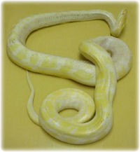
Inclusion body disease is seen in Boidae. Previously the most commonly affected species was Burmese pythons but the disease is now most often seen in Boa constrictors. It appears to be relatively uncommon in royal pythons and has not been reported in Rosy boas. The occurrence of the disease in non-boids cannot be ruled out since typical signs and lesions have been reported in a kingsnake. IBD is primarily seen in adult snakes, however all age groups should be considered susceptible. Young snakes tend to develop an acute infection with a mortality rate approaching 100%. Infected adult snakes often experience the disease in a more chronic and debilitating form.
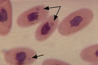
Inclusion body disease (IBD) is a worldwide disease of Boidae and is named for the characteristic intracytoplasmic inclusions seen in the cells of affected snakes. The causative agent of has not been identified although a Retroviridae-like virus has been incriminated.
Examination

Classically pythons developed acute CNS signs and boas presented with regurgitation. In fact clinical signs are very variable among species and individuals and may related to secondary bacterial infections in immunosuppressed boids.
Common clinical signs include the following:
- Lethargy
- Anorexia
- Weight loss
- Lack of weight gain
- Pale oral mucous membranes
- CNS problems
- Incoordination
- Disorientation
- Loss of righting reflexes
- Head tremors
- Opisthotonous
- Paresis to paralysis
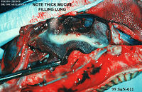
- Regurgitation (partially digested possibly mucous-covered food food about one week post-prandial), stomatitis, diarrhoea, pneumonia, dermatitis and neoplasia.
For more information on physical examination of a snake, see Snake Physical Examination.
Diagnosis
Since IBD presents with a variety of signs, any sick boid without an obvious aetiology should be suspected of IBD. Especially consider those with regurgitation, CNS signs and bacterial infections. A thorough physical examination may rule our other causes. Currently there is no serologic assay available for determining exposure. Diagnostic aids include the following:
Blood
Blood examination may be helpful. Haematology may show a leukocytosis with a lymphocytosis. The white blood cell count is variable, ranging from normal (immune system depression) to as high as 100,000. The leucocytes may show toxic changes. Smears are observed for the presence of inclusion bodies. Typically 1% of cells have the inclusion bodies so it is worthwhile doing a buffy coat smear. Biochemistry may show changes in liver parameters.
Biopsy and histology
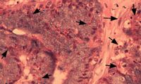
Tissues for biopsy include oesophageal tonsils, liver, kidney and gastric mucosa. The presence of cytoplasmic inclusion bodies on histology is diagnostic for IBD. Sometimes very few inclusions are seen so absence of inclusions does not rule out the disease. For antemortem diagnosis biopsies may have be repeated several times to find inclusions. Characteristic histopathology may be seen in several tissues.
- For more infomation on taking samples from snakes, see Specimen Collection and Evaluation.
Necropsy
Necropsy shows changes associated with secondary bacterial infections. On histology, multiple tissues have been reported to contain cells with inclusions.
- For more information on postmortem of a snake, see Snake Necropsy.
Therapy
There is no known treatment. Euthanasia is recommended.
- For more information on euthanasia, see Lizard and Snake Euthanasia.
Prevention
A sound preventive medicine programme is the protection. All new snakes should be quarantined for a minimum of 90 days before introduction to others. For larger collections a quarantine of 6 months may be more appropriate. The exact route of transmission is unknown so hygiene and mite control are essential. Finally all infected snakes should be euthanased.
For more information on preventative veterinary medicine, see:


