Difference between revisions of "Pancreatitis"
Fiorecastro (talk | contribs) |
|||
| (37 intermediate revisions by 4 users not shown) | |||
| Line 1: | Line 1: | ||
| − | {{ | + | {{unfinished}} |
| − | |||
| − | |||
| − | |||
| − | + | ||
| + | ==Description== | ||
| + | Occurs following activation of digestive enzymes within the [[Pancreas - Anatomy & Physiology|pancreas]] leading to autodigestion of the gland. Can be referred to as Acute or chronic pancreatitis. | ||
| + | Acute Pancreatitis is rapid onset inflammation of the pancreas with little or no pathological changes occuring post recovery. This may completely resolve or 'wax and wane' in the future. | ||
| − | + | Chronic Pancreatitis is continued inflammation leading to irreversible pathological changes (fibrosis, atrophy) and possible decreases in function. | |
| − | The specific cause is usually idiopathic but several risk factors exist including | + | The specific cause is usually idiopathic but several risk factors exist including |
| − | A '''Nutritional''' basis which refers to obesity, low protein and high fat diets, feeding of ethionine and | + | A '''Nutritional''' basis which refers to obesity, low protein and high fat diets, feeding of ethionine, hypertriglyceridaemia and fatty meals. |
'''Drugs and toxins''' including L-asparginase, oestrogen, azathioprine, potassium bromide, furosemide, thiazide diuretics, salicylates, [[Tetracyclines|tetracyclines]], [[Sulphonamides|sulphonamides]], vinca alkaloids, zinc toxicosis, cholinesterase inhibitor insecticides, cholinergic agonist and hypercalcaemia. | '''Drugs and toxins''' including L-asparginase, oestrogen, azathioprine, potassium bromide, furosemide, thiazide diuretics, salicylates, [[Tetracyclines|tetracyclines]], [[Sulphonamides|sulphonamides]], vinca alkaloids, zinc toxicosis, cholinesterase inhibitor insecticides, cholinergic agonist and hypercalcaemia. | ||
| − | '''Pancreatic | + | '''Pancreatic Duct obstruction''' which is caused by biliary calculi, sphincter spasm, duct wall oedema, duodenal wall oedema, neoplasia, parasites, trauma and iatrogenic reasons. |
| − | '''Duodenal juice reflux, | + | '''Duodenal juice reflux, Pancreatic trauma, ischaemia and reperfusion''' which includes duodenal juice reflux into the pancreatic duct, surgical intervention, shock, anaemia, venous occlusion and hypotension. |
'''Other''' risk factors include parasitic (babesiosis), viral, mycoplasmal, end stage renal disease, liver disease and auto-immune diseases. | '''Other''' risk factors include parasitic (babesiosis), viral, mycoplasmal, end stage renal disease, liver disease and auto-immune diseases. | ||
| Line 22: | Line 22: | ||
Cats mainly suffer from mild chronic interstitial pancreatitis. | Cats mainly suffer from mild chronic interstitial pancreatitis. | ||
| + | ==Signalment== | ||
| + | Predisposed breeds include: | ||
| + | <gallery> | ||
| + | Image:Labrador.jpg|'''Labradors''' <br> Ellen Levy Finch (2004) WikiMedia Commons | ||
| + | Image:Miniature Poodle.jpg|'''Miniature Poodles''' <br> Belinda (2005) WikiMedia Commons | ||
| + | Image:Miniature schnauzer.jpg|'''Miniature Schnauzers''' <br> MagnusK (2006) WikiMedia Commons | ||
| + | Image:Yorkshire Terrier.jpg|'''Yorkshire terriers''' <br> Jlcerso (2007) WikiMedia Commons | ||
| + | </gallery> | ||
| − | |||
| − | + | Increased risk of disease occurs with obesity, [[Diabetes Mellitus|diabetes mellitus]], [[Adrenal Glands - Pathology#Adrenal Hyperfunction|hyperadrenocorticalism]], prior [[Alimentary - Anatomy & Physiology|GIT]] disease or epilepsy. | |
| − | + | Additonally middle aged dogs are more commonly affected and Male and speyed females are affected more frequently than entire females. | |
| − | === | + | ==Diagnosis== |
| + | ===History and Clinical Signs=== | ||
| + | There is often a history of eating a fatty meal. | ||
| − | + | Clinical signs include anorexia, vomiting, abdominal pain, lethargy, depression and Nausea. | |
| + | [[Diarrhoea|Diarrhoea]] is also a common feature sometimes with blood, fresh or melaena this occurs due to the proximity of inflamed pancreas to the [[Duodenum - Anatomy & Physiology|duodenum]] and [[Colon - Anatomy & Physiology|colon]]. | ||
| + | More severe cases may present in [[Shock - Pathology|shock]], [[Kidney Renal Failure - Pathology#Acute|acute renal failure]], [[Icterus|jaundiced]] (due to focal hepatic necrosis), or with [[:Category:Altered Impulse Formations|cardiac arrhythmias]]. [[Lungs Circulatory - Pathology#Pulmonary oedema|Pulmonary oedema]], pleural effusions, widespread haemorrhage, [[Disseminated Intravascular Coagulation|DIC]], mild ascites, dehydration (Mild to moderate) and pyrexia may also be present. | ||
| + | Acute haemorrhagic pancreatitis may present as [[Shock - Pathology|shock]] and collapse. | ||
| + | A cranial abdominal mass may be palpated. | ||
| − | + | ||
| + | Affected cats have a very varied presentation. If severe, they present with lethargy and anorexia with vomiting and abdominal pain being reported less than in the dog, hypothermia is also common sign occuring in 68% of affected cats. Mild chronic pancreatitis may show anorexia and weight loss. | ||
| − | + | ===Laboratory Tests=== | |
| + | On Haematology there may be a leucocytosis, an increased PCV due to dehydration, thrombocytopaenia, neutrophilia and a left shift. | ||
| − | + | On Biochemistry changes may include an azotaemia, increased liver enzymes, hyperbilirubinaemia | |
| + | hyperglycaemia in cases of nectrotizing pancreatitis, hypoglycaemia in cats with suppurative pancreatitis | ||
| + | In dogs hypercholesterolaemia and hypertriglyceridaemia are also common changes. | ||
| − | + | An increase in pancreatic digestive enzymes (amylase, lipase, trypsin-like immunoreactivity (TLI), phospholipase A2 and pancreatic lipase immunoreactivity (PLI) will also be present. | |
| − | + | ===Pancreas-specific laboratory tests=== | |
| + | All pancreatic enzymes increase following renal failure (apart from PLI) making it difficult to determine the true cause of the increase. However increases of three fold are mainly due to pancreatitis, whereas five fold increases are rarely not found to be pancreatitis. Rises in lipase, amylase and phospholipase A2 may also be hepatic, gastric, intestinal or neoplastic in origin. | ||
| − | + | '''In cats:''' Amylase and lipase are of no diagnostic value. Serum feline trypsin-like immunoreactivity (fTLI) is a specific test for exocrine pancreatic function but the test's sensitivity varies between 30% and 60%. In comparison, the serum feline pancreatic lipase immunoreactivity test (fPLI) has been found to be more specific and sensitive in diagnosing feline pancreatitis. | |
| − | + | '''In dogs:''' Marked increases in serum lipase is a more reliable marker than amylase. However corticosteroid administration raises lipase activity by up to five fold. Serum canine pancreatic lipase immunoreactivity (cPLI) is the most sensitive and specific test for diagnosing canine pancreatitis. | |
| − | === | + | ===Diagnostic Imaging=== |
| + | '''Survey Radiography''': Rarely helpful but findings may include | ||
| + | In the right cranial abdomen an increased density, decreased contrast, decreased granularity and the stomach may be displaced to the left. | ||
| + | Additionally the descending duodenum may be displaced to the right, with the presence of a medial mass and thickened walls. | ||
| + | Gastric distension may be visible and barium passage may be delayed indicating abnormal peristalsis. | ||
| − | + | Radiography is used to rule out differentials. | |
| − | + | '''Abdominal Ultrasound''' is highly specific with a sensitivity of 70% in dogs and 30% in cats but is operator-dependant. Findings include | |
| + | pancreatic enlargement, peritoneal effusion, hypoechogenic pancreas (pancreatic necrosis) and hyperechogenic surrounding tissue. | ||
| − | + | ===Exploratory Laparotomy/Necropsy Findings=== | |
| + | The pancreas will be oedematous and soft with fibrinous attachments to surrounding organs, there may be free fluid within the peritoneal cavity and pancreas liquefaction if severe enough. | ||
| + | Pseudocysts may be present, as well as omental and pancreatic haemorrhages and areas of fat necrosis. | ||
| − | + | A biopsy should be taken to provide evidence of inflammation. | |
| − | + | ==Treatment== | |
| + | ===Acute Treatment=== | ||
| + | The general treatment involves fluid correction and maintenance while any underlying cause is treated. Support is then given to allow the inflammatory process to subside. Oral feeding should be witheld for a short period in vomiting patients but enteral and parenteral feeding can be well tolerated. | ||
| − | + | Analgesia should always be given even without signs of pain. Recommended options include subcutaneous [[Opioids#Pethidine|pethidine]], intravenous or continuous rate infusion [[Opioids#Morphine|morphine]] or transdermal [[Opioids#Fentanyl|fentanyl]]. Dogs can also be given intraperitoneal [[Local Anaesthetics#Lidocaine|lidocaine]] or [[Local Anaesthetics#Bupivicaine|bupivicaine]]. | |
| − | | | ||
| − | |||
| − | |||
| − | + | If a pancreatic infection is suspected then [[Antibiotics|Antibiotics]] should be administered, [[Potentiated-Sulphonamides|trimethoprim-sulphonamide]] and [[Fluoroquinolones|enrofloxacin]] have good penetration to the pancreas. | |
| − | |||
| − | | | ||
| − | | | ||
| − | | | ||
| − | |||
| − | |||
| − | |||
| − | + | Food can be gradually introduced with a low protein and fat content as these are more likely to cause signs. Fat can be further introduced if symptoms have still not returned. If signs reoccur then further starvation should be carried out. Total parenteral nutrition can be used to sustain animals that are unable to tolerate food at all. | |
| − | + | Cases often require supportive care, aggressive [[Fluid Therapy|fluid therapy]] will be needed to treat dehydration and fluid loss from [[Diarrhoea|diarrhoea]] and [[Control of Feeding - Anatomy & Physiology#The Vomit Reflex|vomiting]]. Renal function and potassium levels should be monitored and if necessary pottasium should be supplemented. | |
| + | Patients may also develop a metabolic acidosis in acute pancreatitis or be alkalotic due to vomiting. Should [[Diabetes Mellitus|diabetes mellitus]] develop, this may require treatment with insulin. Further management may be required for respiratory distress, bleeding disorders, renal failure, cardiovascular problems and neurological disorders. | ||
| − | + | Additionally a whole blood or plasma transfusion can be given with severe disease to replace α-macroglobulins. Albumin also provides oncotic support and limits pancreatic ischaemia and oedema. | |
| − | + | For short term use in fulminating pancreatitis [[Steroids|Corticosteroids]] can be given alongside fluids. Long term treatment may lead to unwanted complications. | |
| − | + | ===Long-term treatment=== | |
| + | In most patients that have one episode, they may only need to avoid fatty foods. Recurrent hypertriglyceridaemia may need pharmacological intervention. | ||
| − | + | ==Prognosis== | |
| + | The disease varies widely and the prognosis can vary from full recovery to death. Generally if the case is an uncomplicated single episode patients will make a good recovery. | ||
| − | + | ==References== | |
| − | |||
| − | |||
| − | |||
| − | |||
| − | |||
| − | |||
| − | |||
| − | |||
| − | |||
| − | |||
| − | |||
| − | |||
| − | |||
| − | |||
| − | |||
| − | |||
| − | |||
| − | |||
| − | |||
| − | |||
| − | |||
| − | + | For further information on canine pancreatitis see: [http://inpractice.bvapublications.com/cgi/reprint/26/2/64?maxtoshow=&HITS=10&hits=10&RESULTFORMAT=&fulltext=feline+pancreatitis&searchid=1&FIRSTINDEX=0&sortspec=relevance&resourcetype=HWCIT Pancreatitis in the dog:. dealing with a spectrum of disease] In Practice article | |
| − | + | For further information on feline pancreatitis see: [http://inpractice.bvapublications.com/cgi/reprint/29/8/470?maxtoshow=&HITS=10&hits=10&RESULTFORMAT=&fulltext=feline+pancreatitis&searchid=1&FIRSTINDEX=0&sortspec=relevance&resourcetype=HWCIT Feline pancreatitis: current concepts and treatment guidelines] In Practice article | |
| + | Hall, E.J, Simpson, J.W. and Williams, D.A. (2005) '''BSAVA Manual of Canine and Feline Gastroenterology (2nd Edition)''' ''BSAVA'' | ||
| − | [[Category: | + | Merck & Co (2008) '''The Merck Veterinary Manual''' |
| + | [[Category:Pancreas_-_Inflammatory_Pathology]][[Category:Dog]][[Category:Cat]] | ||
| + | [[Category:To_Do_-_Caz]] | ||
Revision as of 17:12, 9 August 2010
| This article is still under construction. |
Description
Occurs following activation of digestive enzymes within the pancreas leading to autodigestion of the gland. Can be referred to as Acute or chronic pancreatitis. Acute Pancreatitis is rapid onset inflammation of the pancreas with little or no pathological changes occuring post recovery. This may completely resolve or 'wax and wane' in the future.
Chronic Pancreatitis is continued inflammation leading to irreversible pathological changes (fibrosis, atrophy) and possible decreases in function.
The specific cause is usually idiopathic but several risk factors exist including
A Nutritional basis which refers to obesity, low protein and high fat diets, feeding of ethionine, hypertriglyceridaemia and fatty meals.
Drugs and toxins including L-asparginase, oestrogen, azathioprine, potassium bromide, furosemide, thiazide diuretics, salicylates, tetracyclines, sulphonamides, vinca alkaloids, zinc toxicosis, cholinesterase inhibitor insecticides, cholinergic agonist and hypercalcaemia.
Pancreatic Duct obstruction which is caused by biliary calculi, sphincter spasm, duct wall oedema, duodenal wall oedema, neoplasia, parasites, trauma and iatrogenic reasons.
Duodenal juice reflux, Pancreatic trauma, ischaemia and reperfusion which includes duodenal juice reflux into the pancreatic duct, surgical intervention, shock, anaemia, venous occlusion and hypotension.
Other risk factors include parasitic (babesiosis), viral, mycoplasmal, end stage renal disease, liver disease and auto-immune diseases.
Cats mainly suffer from mild chronic interstitial pancreatitis.
Signalment
Predisposed breeds include:
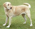
Labradors
Ellen Levy Finch (2004) WikiMedia Commons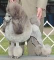
Miniature Poodles
Belinda (2005) WikiMedia Commons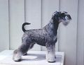
Miniature Schnauzers
MagnusK (2006) WikiMedia Commons
Yorkshire terriers
Jlcerso (2007) WikiMedia Commons


Increased risk of disease occurs with obesity, diabetes mellitus, hyperadrenocorticalism, prior GIT disease or epilepsy.
Additonally middle aged dogs are more commonly affected and Male and speyed females are affected more frequently than entire females.
Diagnosis
History and Clinical Signs
There is often a history of eating a fatty meal.
Clinical signs include anorexia, vomiting, abdominal pain, lethargy, depression and Nausea. Diarrhoea is also a common feature sometimes with blood, fresh or melaena this occurs due to the proximity of inflamed pancreas to the duodenum and colon. More severe cases may present in shock, acute renal failure, jaundiced (due to focal hepatic necrosis), or with cardiac arrhythmias. Pulmonary oedema, pleural effusions, widespread haemorrhage, DIC, mild ascites, dehydration (Mild to moderate) and pyrexia may also be present. Acute haemorrhagic pancreatitis may present as shock and collapse. A cranial abdominal mass may be palpated.
Affected cats have a very varied presentation. If severe, they present with lethargy and anorexia with vomiting and abdominal pain being reported less than in the dog, hypothermia is also common sign occuring in 68% of affected cats. Mild chronic pancreatitis may show anorexia and weight loss.
Laboratory Tests
On Haematology there may be a leucocytosis, an increased PCV due to dehydration, thrombocytopaenia, neutrophilia and a left shift.
On Biochemistry changes may include an azotaemia, increased liver enzymes, hyperbilirubinaemia hyperglycaemia in cases of nectrotizing pancreatitis, hypoglycaemia in cats with suppurative pancreatitis In dogs hypercholesterolaemia and hypertriglyceridaemia are also common changes.
An increase in pancreatic digestive enzymes (amylase, lipase, trypsin-like immunoreactivity (TLI), phospholipase A2 and pancreatic lipase immunoreactivity (PLI) will also be present.
Pancreas-specific laboratory tests
All pancreatic enzymes increase following renal failure (apart from PLI) making it difficult to determine the true cause of the increase. However increases of three fold are mainly due to pancreatitis, whereas five fold increases are rarely not found to be pancreatitis. Rises in lipase, amylase and phospholipase A2 may also be hepatic, gastric, intestinal or neoplastic in origin.
In cats: Amylase and lipase are of no diagnostic value. Serum feline trypsin-like immunoreactivity (fTLI) is a specific test for exocrine pancreatic function but the test's sensitivity varies between 30% and 60%. In comparison, the serum feline pancreatic lipase immunoreactivity test (fPLI) has been found to be more specific and sensitive in diagnosing feline pancreatitis.
In dogs: Marked increases in serum lipase is a more reliable marker than amylase. However corticosteroid administration raises lipase activity by up to five fold. Serum canine pancreatic lipase immunoreactivity (cPLI) is the most sensitive and specific test for diagnosing canine pancreatitis.
Diagnostic Imaging
Survey Radiography: Rarely helpful but findings may include In the right cranial abdomen an increased density, decreased contrast, decreased granularity and the stomach may be displaced to the left. Additionally the descending duodenum may be displaced to the right, with the presence of a medial mass and thickened walls. Gastric distension may be visible and barium passage may be delayed indicating abnormal peristalsis.
Radiography is used to rule out differentials.
Abdominal Ultrasound is highly specific with a sensitivity of 70% in dogs and 30% in cats but is operator-dependant. Findings include pancreatic enlargement, peritoneal effusion, hypoechogenic pancreas (pancreatic necrosis) and hyperechogenic surrounding tissue.
Exploratory Laparotomy/Necropsy Findings
The pancreas will be oedematous and soft with fibrinous attachments to surrounding organs, there may be free fluid within the peritoneal cavity and pancreas liquefaction if severe enough. Pseudocysts may be present, as well as omental and pancreatic haemorrhages and areas of fat necrosis.
A biopsy should be taken to provide evidence of inflammation.
Treatment
Acute Treatment
The general treatment involves fluid correction and maintenance while any underlying cause is treated. Support is then given to allow the inflammatory process to subside. Oral feeding should be witheld for a short period in vomiting patients but enteral and parenteral feeding can be well tolerated.
Analgesia should always be given even without signs of pain. Recommended options include subcutaneous pethidine, intravenous or continuous rate infusion morphine or transdermal fentanyl. Dogs can also be given intraperitoneal lidocaine or bupivicaine.
If a pancreatic infection is suspected then Antibiotics should be administered, trimethoprim-sulphonamide and enrofloxacin have good penetration to the pancreas.
Food can be gradually introduced with a low protein and fat content as these are more likely to cause signs. Fat can be further introduced if symptoms have still not returned. If signs reoccur then further starvation should be carried out. Total parenteral nutrition can be used to sustain animals that are unable to tolerate food at all.
Cases often require supportive care, aggressive fluid therapy will be needed to treat dehydration and fluid loss from diarrhoea and vomiting. Renal function and potassium levels should be monitored and if necessary pottasium should be supplemented. Patients may also develop a metabolic acidosis in acute pancreatitis or be alkalotic due to vomiting. Should diabetes mellitus develop, this may require treatment with insulin. Further management may be required for respiratory distress, bleeding disorders, renal failure, cardiovascular problems and neurological disorders.
Additionally a whole blood or plasma transfusion can be given with severe disease to replace α-macroglobulins. Albumin also provides oncotic support and limits pancreatic ischaemia and oedema.
For short term use in fulminating pancreatitis Corticosteroids can be given alongside fluids. Long term treatment may lead to unwanted complications.
Long-term treatment
In most patients that have one episode, they may only need to avoid fatty foods. Recurrent hypertriglyceridaemia may need pharmacological intervention.
Prognosis
The disease varies widely and the prognosis can vary from full recovery to death. Generally if the case is an uncomplicated single episode patients will make a good recovery.
References
For further information on canine pancreatitis see: Pancreatitis in the dog:. dealing with a spectrum of disease In Practice article
For further information on feline pancreatitis see: Feline pancreatitis: current concepts and treatment guidelines In Practice article
Hall, E.J, Simpson, J.W. and Williams, D.A. (2005) BSAVA Manual of Canine and Feline Gastroenterology (2nd Edition) BSAVA
Merck & Co (2008) The Merck Veterinary Manual