Difference between revisions of "Avian Female Reproductive System"
Jump to navigation
Jump to search
m (Text replace - "-_Anatomy_%26_Physiology" to "- Anatomy & Physiology") |
|||
| (12 intermediate revisions by the same user not shown) | |||
| Line 1: | Line 1: | ||
| + | {{toplink | ||
| + | |backcolour =EED2EE | ||
| + | |linkpage =Reproductive System - Anatomy & Physiology | ||
| + | |linktext =Reproductive System | ||
| + | |maplink = Reproductive System (Content Map) - Anatomy & Physiology | ||
| + | |sublink1=Exotics - Avian Reproductive Anatomy and Physiology - Anatomy & Physiology | ||
| + | |subtext1=AVIAN REPRODUCTIVE | ||
| + | }} | ||
| + | <br> | ||
== Ovary == | == Ovary == | ||
In the first 5 months after hatching, the ovary gradually develops from a small irregular structure with a finely granular surface into one where individual follicles can be observed. | In the first 5 months after hatching, the ovary gradually develops from a small irregular structure with a finely granular surface into one where individual follicles can be observed. | ||
* Most birds have only left ovary but 2 ovaries are typical of many raptors. | * Most birds have only left ovary but 2 ovaries are typical of many raptors. | ||
| − | * '''Left Ovary''' lies caudal to the [[Adrenal_Glands_- Anatomy & Physiology|Adrenal Gland]], close to the cranial tip of the [[Renal Anatomy - Anatomy & Physiology#Common Anatomy|Kidney]]. | + | * '''Left Ovary''' lies caudal to the [[Adrenal_Glands_- Anatomy & Physiology|Adrenal Gland]], close to the cranial tip of the [[Macroscopic Renal Anatomy - Anatomy & Physiology#Common Anatomy|Kidney]]. |
* Consists of: | * Consists of: | ||
** '''Medulla''': vascular with nerve fibres and smooth muscle | ** '''Medulla''': vascular with nerve fibres and smooth muscle | ||
| Line 9: | Line 18: | ||
* Suspended by the '''mesovarium'''. | * Suspended by the '''mesovarium'''. | ||
* Blood supply from the '''cranial renal artery''' | * Blood supply from the '''cranial renal artery''' | ||
| − | ** Artery is very short, making ovariectomy very difficult with risk of [[Haemorrhage|haemorrhage]]. | + | ** Artery is very short, making ovariectomy very difficult with risk of [[Haemorrhage - Pathology|haemorrhage]]. |
| − | * Contains from 500 to several thousand primary [[ | + | * Contains from 500 to several thousand primary [[The_Ovary_-_Oocytes_- Anatomy & Physiology|oocytes]]. |
| − | * [[ | + | * [[The_Ovary_-_Follicles_- Anatomy & Physiology|Follicles]] are '''pedunculated''', suspended by a stalk containing smooth muscle that has a rich vascular and nerve supply. |
| − | ** Large, pendulous [[ | + | ** Large, pendulous [[The_Ovary_-_Follicles_- Anatomy & Physiology|Follicles]] may contact the [[Avian_Digestive_Tract_- Anatomy & Physiology|stomach]], [[Spleen_- Anatomy & Physiology|spleen]] and [[Avian_Intestines_- Anatomy & Physiology|intestines]]. |
** Each consists of a large, yolk-filled oocyte surrounded by a highly vascular follicular wall. | ** Each consists of a large, yolk-filled oocyte surrounded by a highly vascular follicular wall. | ||
* Shortly before [[Exotics_-_Breeding%2C_Ovulation_and_Oviposition_- Anatomy & Physiology#Ovulation|ovulation]], a devascularized white band (stigma) appears opposite the stalk, indicating where the wall will rupture at [[Exotics_-_Breeding%2C_Ovulation_and_Oviposition_- Anatomy & Physiology#Ovulation|ovulation]]. | * Shortly before [[Exotics_-_Breeding%2C_Ovulation_and_Oviposition_- Anatomy & Physiology#Ovulation|ovulation]], a devascularized white band (stigma) appears opposite the stalk, indicating where the wall will rupture at [[Exotics_-_Breeding%2C_Ovulation_and_Oviposition_- Anatomy & Physiology#Ovulation|ovulation]]. | ||
* The empty follicle (calix) regresses following [[Exotics_-_Breeding%2C_Ovulation_and_Oviposition_- Anatomy & Physiology#Ovulation|ovulation]] and disappears in a few days. | * The empty follicle (calix) regresses following [[Exotics_-_Breeding%2C_Ovulation_and_Oviposition_- Anatomy & Physiology#Ovulation|ovulation]] and disappears in a few days. | ||
| − | * No [[ | + | * No [[The_Ovary_-Corpus_Luteum_- Anatomy & Physiology|Corpus Luteum]] is required, since there is no embryo to maintain as there is in mammals. |
== Oviduct == | == Oviduct == | ||
| Line 32: | Line 41: | ||
* Occupies the left,dorsal part of the body cavity. | * Occupies the left,dorsal part of the body cavity. | ||
| − | * Related to the [[Renal Anatomy - Anatomy & Physiology#Common Anatomy|Kidney]], [[Avian_Intestines_- Anatomy & Physiology|intestines]] and [[Gizzard_- Anatomy & Physiology|gizzard]]. | + | * Related to the [[Macroscopic Renal Anatomy - Anatomy & Physiology#Common Anatomy|Kidney]], [[Avian_Intestines_- Anatomy & Physiology|intestines]] and [[Gizzard_- Anatomy & Physiology|gizzard]]. |
* Massive coiled structure approximately twice body length when fully functional.. | * Massive coiled structure approximately twice body length when fully functional.. | ||
** Much reduced in juveniles and during the non-laying period. | ** Much reduced in juveniles and during the non-laying period. | ||
| Line 69: | Line 78: | ||
* Subsequent to [[Exotics_-_Breeding%2C_Ovulation_and_Oviposition_- Anatomy & Physiology#Ovulation|ovulation]], the ovum is engulfed by the Infundibulum. | * Subsequent to [[Exotics_-_Breeding%2C_Ovulation_and_Oviposition_- Anatomy & Physiology#Ovulation|ovulation]], the ovum is engulfed by the Infundibulum. | ||
* Ovum resides here for '''15-30 minutes'''. | * Ovum resides here for '''15-30 minutes'''. | ||
| − | * '''Fertilization''' must take place in the funnel section of the Infundibulum before the [[ | + | * '''Fertilization''' must take place in the funnel section of the Infundibulum before the [[The_Ovary_-_Oocytes_- Anatomy & Physiology|oocyte]] gets surrounded by [[Exotics_-_Composition_and_Formation_of_the_Egg_- Anatomy & Physiology#Albumin|albumin]]. |
* Penetration by sperm usually occurs within 15 minutes of [[Exotics_-_Breeding%2C_Ovulation_and_Oviposition_- Anatomy & Physiology#Ovulation|ovulation]]. | * Penetration by sperm usually occurs within 15 minutes of [[Exotics_-_Breeding%2C_Ovulation_and_Oviposition_- Anatomy & Physiology#Ovulation|ovulation]]. | ||
| − | * Infundibular glands provide the chalaziferous layer, the thin coating of dense [[ | + | * Infundibular glands provide the chalaziferous layer, the thin coating of dense [[Exotics_-_Composition_and_Formation_of_the_Egg_- Anatomy & Physiology#Albumin|albumin]] around the [[Exotics_-_Composition_and_Formation_of_the_Egg_- Anatomy & Physiology#Yolk|yolk]]. |
** Chalazae, the coiled strands that suspend the yolk and allow it to rotate so that the ferminal disc remains uppermost, develop further along the genital duct. | ** Chalazae, the coiled strands that suspend the yolk and allow it to rotate so that the ferminal disc remains uppermost, develop further along the genital duct. | ||
| − | * Some species have a [[ | + | * Some species have a [[Exotics_-_Anatomy_of_the_Female_Reproductive_System_- Anatomy & Physiology#Sperm_Host_Glands|'''sperm host gland''']] in this area to store sperm for later fertilization. |
=== Magnum === | === Magnum === | ||
| Line 90: | Line 99: | ||
* Demarcated from the Magnum by a narrow aglandular zone. | * Demarcated from the Magnum by a narrow aglandular zone. | ||
| − | * Folds less prominent than the Magnum, but the glands secrete more [[ | + | * Folds less prominent than the Magnum, but the glands secrete more [[Exotics_-_Composition_and_Formation_of_the_Egg_- Anatomy & Physiology#Albumin|albumin]]. |
* Divides the Magnum from the Uterus. | * Divides the Magnum from the Uterus. | ||
* Present in '''poultry''' but not in Psittacines. | * Present in '''poultry''' but not in Psittacines. | ||
* Ovum resides here for '''~1-2 hours''' | * Ovum resides here for '''~1-2 hours''' | ||
| − | * Inner and outer [[ | + | * Inner and outer [[Exotics_-_Composition_and_Formation_of_the_Egg_- Anatomy & Physiology#Shell|'''shell membrane formation''']]. |
=== Uterus === | === Uterus === | ||
| − | * Holds the egg during [[ | + | * Holds the egg during [[Exotics_-_Composition_and_Formation_of_the_Egg_- Anatomy & Physiology#Shell|shell formation]]. |
* 80% of time is spent here. | * 80% of time is spent here. | ||
* Very vascular to aid calcium deposition. | * Very vascular to aid calcium deposition. | ||
| Line 132: | Line 141: | ||
| − | [[Category: | + | [[Category:Reproductive System]] |
| − | |||
Revision as of 15:21, 29 November 2010
|
|
Ovary
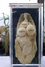
In the first 5 months after hatching, the ovary gradually develops from a small irregular structure with a finely granular surface into one where individual follicles can be observed.
- Most birds have only left ovary but 2 ovaries are typical of many raptors.
- Left Ovary lies caudal to the Adrenal Gland, close to the cranial tip of the Kidney.
- Consists of:
- Medulla: vascular with nerve fibres and smooth muscle
- Cortex: peripheral
- Suspended by the mesovarium.
- Blood supply from the cranial renal artery
- Artery is very short, making ovariectomy very difficult with risk of haemorrhage.
- Contains from 500 to several thousand primary oocytes.
- Follicles are pedunculated, suspended by a stalk containing smooth muscle that has a rich vascular and nerve supply.
- Large, pendulous Follicles may contact the stomach, spleen and intestines.
- Each consists of a large, yolk-filled oocyte surrounded by a highly vascular follicular wall.
- Shortly before ovulation, a devascularized white band (stigma) appears opposite the stalk, indicating where the wall will rupture at ovulation.
- The empty follicle (calix) regresses following ovulation and disappears in a few days.
- No Corpus Luteum is required, since there is no embryo to maintain as there is in mammals.
Oviduct
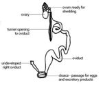
The oviduct may be divided into the infundibulum, magnum, isthmus, uterus and vagina. The oviduct not only conducts the fertilized ovum to the Cloaca, but adds substantial amounts of nutrients. It also encloses the ovum within membranes and a shell, providing protection for the embryo. It conveys spermatozoa to the ovum for immediate fertilization and stores them for future use. One insemination is sufficient to fertilize the ova released during the following 10 days.
- The right oviduct regresses in development under the influence of MIH.
- In most birds only the left oviduct is present and functional. The right oviduct can be identified as a strand of tissue on the right side along the ventral side of the caudal vena cava.
Anatomical Structure and Location
- Occupies the left,dorsal part of the body cavity.
- Related to the Kidney, intestines and gizzard.
- Massive coiled structure approximately twice body length when fully functional..
- Much reduced in juveniles and during the non-laying period.
- Suspended from the roof of the body cavity by a peritoneal fold (mesoviductus).
- Some coils are connected by a ventral continuation that forms the muscular ventral ligament.
Blood Flow and Innervation
- Blood flow to shell gland increased during the presence of a calcifying egg
- Innervated by both sympathetic and parasympathetic nerves
- Infundibulum is via the aortic plexus.
- Magnum is by the aortic and renal plexuses.
- Sympathetic innervation of the shell gland (Vagina) is via the hypogastric nerve, a direct continuation of the aortic plexus.
Oviduct Motility
- Cilia are found along the entire length of the Oviduct. These facillitate sperm transport.
- Egg transported by contractions of the Oviduct musculature.
Histological Structure
- Serosa
- Tunica Muscularis
- Outer spiral layer
- Inner circular layer
- Scanty submucosa
- Tunica Mucosa contains many glands
Infundibulum
- Consists of a fluted and tubular part.
- Fluted part is thin-walled and stretched to form a slit (Infundibular Ostium).
- Lateral end is attached to the body wall near the last rib.
- Ostium is positioned by the left abdominal air sac in such a way that it can grasp the newly released oocyte.
- Fluted part is thin-walled and stretched to form a slit (Infundibular Ostium).
- Subsequent to ovulation, the ovum is engulfed by the Infundibulum.
- Ovum resides here for 15-30 minutes.
- Fertilization must take place in the funnel section of the Infundibulum before the oocyte gets surrounded by albumin.
- Penetration by sperm usually occurs within 15 minutes of ovulation.
- Infundibular glands provide the chalaziferous layer, the thin coating of dense albumin around the yolk.
- Chalazae, the coiled strands that suspend the yolk and allow it to rotate so that the ferminal disc remains uppermost, develop further along the genital duct.
- Some species have a sperm host gland in this area to store sperm for later fertilization.
Magnum
- Longest part of the Oviduct.
- Ovum resides here for ~2-3 hours.
- Coiled with numerous tubular glands, giving it a thickened appearance.
- Oestrogen stimulates epithelial stem cells to develop into three morphologically different cell types:
- Tubular cell glands: Produce ovalbumin,lysozyme and conalbumin under the influence of Oestrogen. This gives the lumen a milky-white colour.
- Ciliated cells
- Goblet cells: synthesize avidin following exposure to Progesterone and Oestrogen.
- Oestrogen stimulates epithelial stem cells to develop into three morphologically different cell types:
- Calcium, sodium and magnesium are added.
Isthmus
- Demarcated from the Magnum by a narrow aglandular zone.
- Folds less prominent than the Magnum, but the glands secrete more albumin.
- Divides the Magnum from the Uterus.
- Present in poultry but not in Psittacines.
- Ovum resides here for ~1-2 hours
- Inner and outer shell membrane formation.
Uterus
- Holds the egg during shell formation.
- 80% of time is spent here.
- Very vascular to aid calcium deposition.
- Calcification first occurs slowly, increases, then decreases again ~2 hours prior to oviposition.
- Shell pigments (primarily protoporphyrin and biliverdin) are deposited via ciliated cells over 3 hours until 30 minutes before oviposition.
Vagina
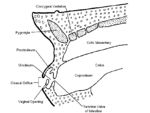
- Muscular S-shaped tube through which the completed egg passes when it is expelled.
- Vagina is separated from the Uterus by a vaginal sphincter which terminates at the Cloaca.
- Ends at a slitlike opening in the lateral wall of the Urodeum.
- Smooth muscle more powerful than the rest of the oviduct.
- Numerous mucosal folds lined by ciliated and non-ciliated cells.
- Few tubular mucosal glands with secretory function.
- No role in egg formation.
- In some species, egg can remain here for hardening before passing out of the oviduct to the Urodeum.
Sperm Host Glands
- Sperm host glands are at the uterovaginal junction of domestic fowl.
- Sperm can be stored here and remain viable for 7-14 days in the Chicken or over 21 days in the Turkey.
- No innervation or contractile tissue.
- Well-developed vascular system.
Cloacal Urodeum
- Urodeum is the middle compartment of the Cloaca.
- Separated from other parts by circular mucosal folds.
- Ureters and genital ducts empty into its dorsal wall.
- Left Oviduct opens into a small mound, which is covered by a small membrane in ducks,geese and swans until sexual maturity.


